

3D airway geometry analysis of factors in airway navigation failure for lung nodules
- PMID: 38965621
- PMCID: PMC11223435
- DOI: 10.1186/s40644-024-00730-7
3D airway geometry analysis of factors in airway navigation failure for lung nodules
Abstract
Background: This study aimed to quantitatively reveal contributing factors to airway navigation failure during radial probe endobronchial ultrasound (R-EBUS) by using geometric analysis in a three-dimensional (3D) space and to investigate the clinical feasibility of prediction models for airway navigation failure.
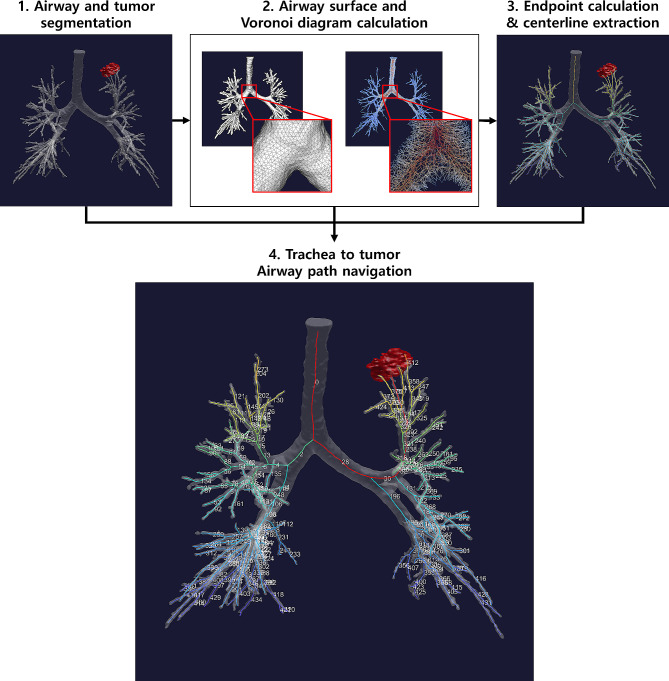
Methods: We retrospectively reviewed patients who underwent R-EBUS between January 2017 and December 2018. Geometric quantification was analyzed using in-house software built with open-source python libraries including the Vascular Modeling Toolkit ( http://www.vmtk.org ), simple insight toolkit ( https://sitk.org ), and sci-kit image ( https://scikit-image.org ). We used a machine learning-based approach to explore the utility of these significant factors.
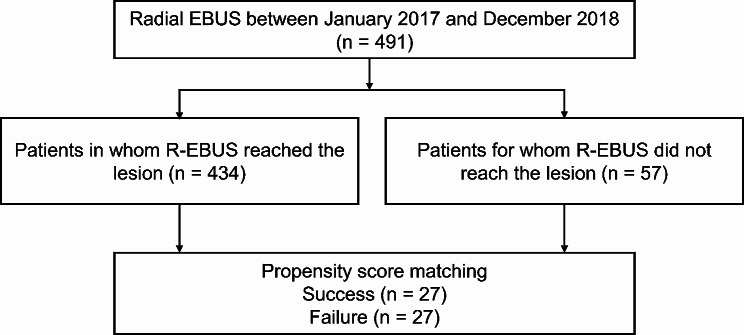
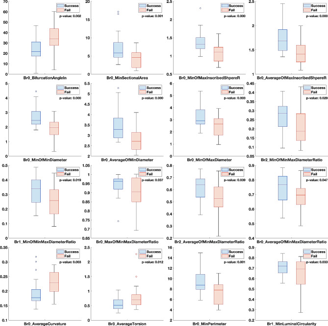
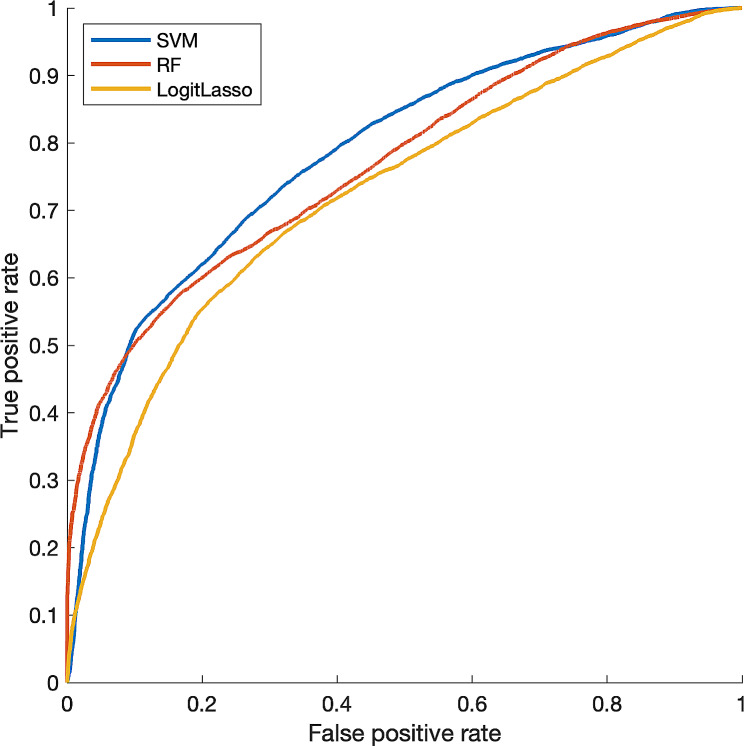
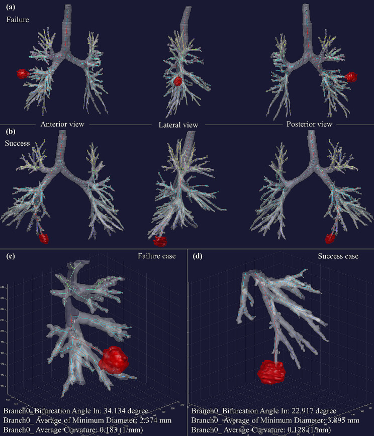
Results: Of the 491 patients who were eligible for analysis (mean age, 65 years +/- 11 [standard deviation]; 274 men), the target lesion was reached in 434 and was not reached in 57. Twenty-seven patients in the failure group were matched with 27 patients in the success group based on propensity scores. Bifurcation angle at the target branch, the least diameter of the last section, and the curvature of the last section are the most significant and stable factors for airway navigation failure. The support vector machine can predict airway navigation failure with an average area under the curve of 0.803.
Conclusions: Geometric analysis in 3D space revealed that a large bifurcation angle and a narrow and tortuous structure of the closest bronchus from the lesion are associated with airway navigation failure during R-EBUS. The models developed using quantitative computer tomography scan imaging show the potential to predict airway navigation failure.
Keywords: Bronchoscopy; Solitary pulmonary nodule; Tomography scanners; X-Ray computed.
© 2024. The Author(s).
Conflict of interest statement
The authors of this manuscript declare no relationships with any companies, whose products or services may be related to the subject matter of the article.
Figures
Similar articles
-
Diagnosis of lung nodules with peripheral/radial endobronchial ultrasound-guided transbronchial biopsy.J Bronchology Interv Pulmonol. 2012 Jan;19(1):5-11. doi: 10.1097/LBR.0b013e31823fcf11. J Bronchology Interv Pulmonol. 2012. PMID: 23207256
-
[Combination of CT mulitplane 3D reconstruction, radial endobronchial ultrasound and rapid on-site evaluation for diagnosing peripheral solitary pulmonary nodules].Zhonghua Yi Xue Za Zhi. 2019 Jan 8;99(2):93-98. doi: 10.3760/cma.j.issn.0376-2491.2019.02.004. Zhonghua Yi Xue Za Zhi. 2019. PMID: 30669745 Chinese.
-
Diagnostic utility of endobronchial ultrasound with a guide sheath under the computed tomography workstation (ziostation) for small peripheral pulmonary lesions.Clin Respir J. 2017 Mar;11(2):185-192. doi: 10.1111/crj.12321. Epub 2015 Jul 2. Clin Respir J. 2017. PMID: 26072931
-
The Diagnostic Accuracy and Sensitivity for Malignancy of Radial-Endobronchial Ultrasound and Electromagnetic Navigation Bronchoscopy for Sampling of Peripheral Pulmonary Lesions: Systematic Review and Meta-analysis.J Bronchology Interv Pulmonol. 2020 Apr;27(2):106-121. doi: 10.1097/LBR.0000000000000645. J Bronchology Interv Pulmonol. 2020. PMID: 31985505
-
Sensitivity of Radial Endobronchial Ultrasound-Guided Bronchoscopy for Lung Cancer in Patients With Peripheral Pulmonary Lesions: An Updated Meta-analysis.Chest. 2020 Apr;157(4):994-1011. doi: 10.1016/j.chest.2019.10.042. Epub 2019 Nov 15. Chest. 2020. PMID: 31738928
Cited by
-
Evaluating diagnostic yield and accuracy as key performance metrics in pulmonary lung lesions.Front Med (Lausanne). 2025 May 7;12:1572779. doi: 10.3389/fmed.2025.1572779. eCollection 2025. Front Med (Lausanne). 2025. PMID: 40400632 Free PMC article.
References
MeSH terms
Grants and funding
- NRF-2022R1A2C1003999/National Research Foundation of Korea (NRF) grant funded by the Korean government (MSIT)
- NRF-2021R1A4A5032806/National Research Foundation of Korea (NRF) grant funded by the Korean government (MSIT)
- SMO1230071/Future Medicine 20*30 Project of the Samsung Medical Center
- 2021-0-02068/Institute of Information & Communications Technology Planning & Evaluation (IITP) grant funded by the Korean government (MSIT)
LinkOut - more resources
Full Text Sources
Medical
