. 2024 Nov;11(42):e2401415.
doi: 10.1002/advs.202401415. Epub 2024 Jul 4.

Modeling the Effects of Protracted Cosmic Radiation in a Human Organ-on-Chip Platform

Affiliations

Modeling the Effects of Protracted Cosmic Radiation in a Human Organ-on-Chip Platform

Daniel Naveed Tavakol et al. Adv Sci (Weinh). 2024 Nov.

Abstract

Galactic cosmic radiation (GCR) is one of the most serious risks posed to astronauts during missions to the Moon and Mars. Experimental models capable of recapitulating human physiology are critical to understanding the effects of radiation on human organs and developing radioprotective measures against space travel exposures. The effects of systemic radiation are studied using a multi-organ-on-a-chip (multi-OoC) platform containing engineered tissue models of human bone marrow (site of hematopoiesis and acute radiation damage), cardiac muscle (site of chronic radiation damage) and liver (site of metabolism), linked by vascular circulation with an endothelial barrier separating individual tissue chambers from the vascular perfusate. Following protracted neutron radiation, the most damaging radiation component in deep space, a greater deviation of tissue function is observed as compared to the same cumulative dose delivered acutely. Further, by characterizing engineered bone marrow (eBM)-derived immune cells in circulation, 58 unique genes specific to the effects of protracted neutron dosing are identified, as compared to acutely irradiated and healthy tissues. It propose that this bioengineered platform allows studies of human responses to extended radiation exposure in an "astronaut-on-a-chip" model that can inform measures for mitigating cosmic radiation injury.

Keywords: bone marrow; heart; human stem cells; liver; mars mission; organ‐on‐chip; radiation; tissue engineering.


Conflict of interest statement

The authors declare no conflict of interest.

Figures
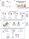
Figure 1
Figure 1
Overall approach. A) Differentiation of human iPSCs into tissue‐specific cells or sourcing of primary cells. B) Integrated, multi‐OoC platform with individual human tissue compartments, vascular flow, and endothelial barriers separating tissues from the flow.[ 19 ] C) Individual tissue formation and maturation over a period of 4–6 weeks, followed by D) integration into the multi‐OoC platforms and exposure to an acute or protracted dose of 0.5 Gy of neutrons over two weeks at Columbia's Radiological Research Facility at Nevis Laboratories (see Figure S1 and Methods). E) End‐point assays performed to characterize the functional, structural, and molecular tissue phenotypes.
Figure 2
Figure 2
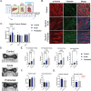
Functional and structural changes in eBMs 14 days after exposure to neutron radiation. A) Schematic of the platform with eBM tissue being characterized. B) Representative flow cytometric gating for CD11b+ cells in the eBM tissue compartment. C) Analysis of hematopoietic progenitors (CD34+, CD34+CD38‐CD90‐CD45RA+ MLPs, CD34+CD38‐CD90‐CD45RA‐ MPPs, CD34+CD38+ CD45RA‐ CMPs/MEPs, and CD34+CD38+ CD45RA+ GMPs) in multi‐OoC after exposures to acute (blue) or protracted (grey) neutrons, in comparison to healthy controls (black). D) Characterization of myeloid cells in eBM compartment (CD11b+ myeloid progeny, CD14+ monocytes, CD11c+CD14+ dendritic cells, CD11b+CD15+CD14‐ granulocytes, and CD11b+CD14+CD15+ M‐MDSCs). Significance was determined by one‐way ANOVA with Tukey's multiple comparisons test (p‐values not shown on plots are p > 0.1). E) Histological staining of eBMs using H&E, Picrosirius Red, and immunostaining for CD45.
Figure 3
Figure 3
Functional and structural changes to eCTs 14 days after exposure to neutron radiation. A) Schematic of the platform with eCT tissue being characterized. B) Histological staining of eCTs using a canonical marker for cardiomyocytes (a‐actinin) and fibroblasts (vimentin). C) Cardiac troponin T (cTnT) concentration in the supernatant of eCT compartments over the duration of the experiment, normalized to control groups, with significance shown by two‐way ANOVA with multiple comparisons. D) Representative bright field image of eCTs at endpoint. E–G) Characterization of contractile function E), tissue morphology at rest F) at the 14‐day endpoint. Each tissue is normalized to itself at baseline and then normalized to the average value of the control group. G) Characterization of excitability of cardiac muscle tissues, normalized to the average value of the control group. Statistical significance was determined by one‐way ANOVA with Welch's correction and corrections for multiple comparisons (C,E,F) or by the Student's t‐test (G) (p‐values not shown on plots are p > 0.1).
Figure 4
Figure 4
Functional and structural changes to eLivs after neutron exposures. A) Schematic of the platform with eLiv tissue being characterized. B) Histological staining of eLivs using canonical markers for hepatocytes (CYP450, CK18), fibroblasts (vimentin), and matrix deposition (COL1A1). C) Albumin and D) urea secretion in eLiv culture supernatant, normalized to the untreated controls. E) Cytokine secretion by eLivs 24 h, 1 week, and 2 weeks post initial radiation exposure, normalized to the untreated controls. Significance was noted by two‐way ANOVAs with multiple comparisons.
Figure 5
Figure 5
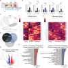
Transcriptomic changes in eBM‐produced circulating immune cells in response to protracted radiation exposure. A) Schematic of the platform with circulating cells being characterized. B) Secreted inflammatory cytokines in the circulatory compartment 24 h post‐irradiation. Statistical significance was performed via one‐way ANOVA without correction for multiple comparisons. C) Principal component analysis (PCA) of control, acute, and protracted circulatory cells gene expression. D) Differentially expressed genes (DEGs) in immune cells in the control versus acute group, control versus protracted group, and acute versus protracted group. E) Volcano plot of significant DEGs in protracted group as compared to healthy controls. F, G) Heatmaps of common radiation damage F) and inflammatory genes (G). H) Gene ontology (GO) pathway analysis of biological processes for upregulated (left) and downregulated (right) processes for protracted versus control groups.
Figure 6
Figure 6
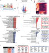
Transcriptomic changes in circulating immune cells following protracted radiation exposure. A) Schematic of the platform with circulating cells being characterized. B) Volcano plot of significant DEGs in the protracted radiation group as compared to the acute radiation group. C) Heatmap of top DEGs. D) GO pathway analysis of upregulated (top) and downregulated (bottom) biological processes following exposure to protracted as compared to acute radiation. E) GO analysis of Wiki Pathways resulting in upregulated (top) and downregulated (bottom) genes following protracted versus acute radiation, with identification of target genes enriched in each pathway. F) Identification of genes specifically responding to protracted radiation exposure. G) List of genes with increased (left, red) and decreased (right, blue) fold changes following exposure to protracted radiation, as compared to either acute radiation or healthy controls. Known radiation response genes are shown in bold.

References

    1. Patel Z. S., Brunstetter T. J., Tarver W. J., Whitmire A. M., Zwart S. R., Smith S. M., Huff J. L., NPJ Microgravity 2020, 6, 33. - PMC - PubMed
    1. Bokhari R. S., Beheshti A., Blutt S. E., Bowles D. E., Brenner D., Britton R., Bronk L., Cao X.u, Chatterjee A., Clay D. E., Courtney C., Fox D. T., Gaber M. W., Gerecht S., Grabham P., Grosshans D., Guan F., Jezuit E. A., Kirsch D. G., Liu Z., Maletic‐Savatic M., Miller K. M., Montague R. A., Nagpal P., Osenberg S., Parkitny L., Pierce N. A., Porada C., Rosenberg S. M., Sargunas P., et al., Life Sci. Space Res. 2022, 35, 105. - PubMed
    1. Krestinina L. Y., Preston D. L., Ostroumova E. V., Degteva M. O., Ron E., Vyushkova O. V., Startsev N. V., Kossenko M. M., Akleyev A. V., Radiat. Res. 2005, 164, 602. - PubMed
    1. Leuraud K., Richardson D. B., Cardis E., Daniels R. D., Gillies M., O'Hagan J. A., Hamra G. B., Haylock R., Laurier D., Moissonnier M., Schubauer‐Berigan M. K., Thierry‐Chef I., Kesminiene A., Lancet Haematol. 2015, 2, e276. - PMC - PubMed
    1. Holden S., Perez R., Hall R., Fallgren C. M., Ponnaiya B., Garty G., Brenner D. J., Weil M. M., Raber J., Radiat. Res. 2021, 196, 31. - PMC - PubMed

LinkOut - more resources