Review
. 2024 Sep 1;11(Pt 5):664-674.
doi: 10.1107/S2052252524005360.

Many locks to one key: N-acetylneuraminic acid binding to proteins

Affiliations
Review

Many locks to one key: N-acetylneuraminic acid binding to proteins

KanagaVijayan Dhanabalan et al. IUCrJ. .

Abstract

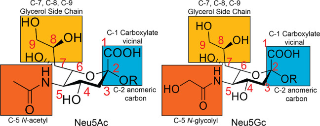
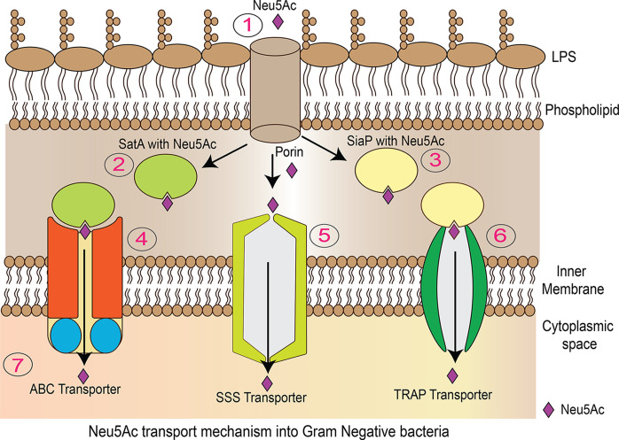
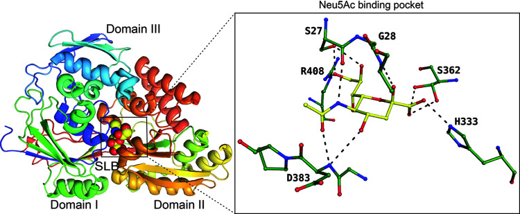
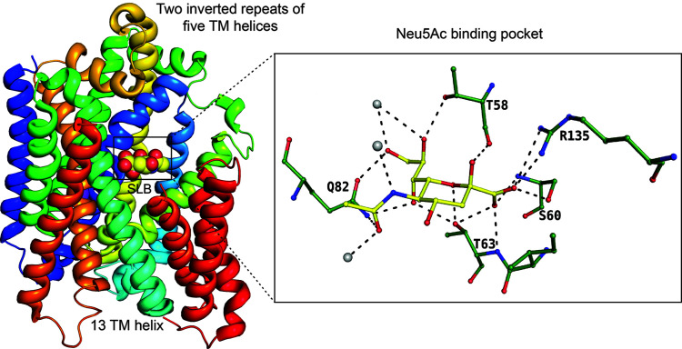
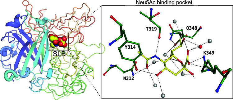
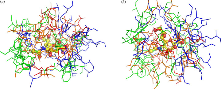
Sialic acids play crucial roles in cell surface glycans of both eukaryotic and prokaryotic organisms, mediating various biological processes, including cell-cell interactions, development, immune response, oncogenesis and host-pathogen interactions. This review focuses on the β-anomeric form of N-acetylneuraminic acid (Neu5Ac), particularly its binding affinity towards various proteins, as elucidated by solved protein structures. Specifically, we delve into the binding mechanisms of Neu5Ac to proteins involved in sequestering and transporting Neu5Ac in Gram-negative bacteria, with implications for drug design targeting these proteins as antimicrobial agents. Unlike the initial assumptions, structural analyses revealed significant variability in the Neu5Ac binding pockets among proteins, indicating diverse evolutionary origins and binding modes. By comparing these findings with existing structures from other systems, we can effectively highlight the intricate relationship between protein structure and Neu5Ac recognition, emphasizing the need for tailored drug design strategies to inhibit Neu5Ac-binding proteins across bacterial species.

Keywords: Gram-negative bacteria; N-acetyl neuraminic acid; Neu5Ac; binding sites; drug discovery; molecular recognition; protein structures; sialic acids.


Figures

Figure 1
Figure 1
Structures of the two most common forms of sialic acid: Neu5Ac and Neu5Gc.
Figure 2
Figure 2
Neu5Ac transport mechanism in Gram-negative bacteria. (1) Porins located in the outer membrane facilitate the transport of Neu5Ac into the periplasmic space. (2) and (4) SatA proteins bind to Neu5Ac molecules and are then associated with the ABC transporter. SiaP proteins (3) also bind to Neu5Ac and assist TRAP transporters (6) in translocating Neu5Ac into the cytoplasm. (5) The SSS transporter facilitates the movement of cargo molecules across the membrane using the energy provided by an ion gradient.
Figure 3
Figure 3
SatA binding to the β-anomer of Neu5Ac (SLB), represented by spheres. The insert displays the binding pocket of SatA and the residues interacting with Neu5Ac (PDB entry 5z99; Gangi Setty et al., 2018 ▸), represented as a ball-and-stick diagram.
Figure 4
Figure 4
SSS transporters binding to the β-anomer of Neu5Ac, represented by spheres. The ball-and-stick diagram displays residues interacting with Neu5Ac (PDB entry 5nva; Wahlgren et al., 2018 ▸). Solvent molecules are shown as grey spheres.
Figure 5
Figure 5
SiaP binding to the β-anomer of Neu5Ac, represented by spheres. As a ball-and-stick diagram, the insert shows the binding site with the residues involved in the interaction with Neu5Ac (PDB entry 4mmp; Gangi Setty et al., 2014 ▸). Solvent atoms are represented as grey spheres.
Figure 6
Figure 6
Illustration of the Neu5Ac utilization pathway. (1) Various transporter proteins facilitate the movement of Neu5Ac from the periplasmic space into the cytoplasm. (2) In the catabolic pathway, the first enzyme, NanA (Neu5Ac lyase), initiates the utilization of Neu5Ac. (3) SiaB (CMP–Neu5Ac synthetase) adds CMP to Neu5Ac in the incorporation pathway. (4) Lic3A (sialyltransferase) incorporates Neu5Ac into the LPS.
Figure 7
Figure 7
NanR binding to the β-anomer of Neu5Ac, represented by spheres. The insert displays the binding site of NanR, showing the residues involved in the interaction with Neu5Ac (PDB entry 6on4; Kalivoda et al., 2013 ▸) as a ball-and-stick diagram. Solvent atoms are represented as grey spheres and the zinc atom as a purple sphere.
Figure 8
Figure 8
NanA binding to the Neu5Gc, represented as spheres. The insert displays the active site of NanA and the residues involved in the interaction with Neu5Gc (PDB entry 4img; Huynh et al., 2013 ▸) as a ball-and-stick diagram. Note that the sugar is the linear form needed for the lysis of the pyruvate moiety. The solvent atom is shown in grey.
Figure 9
Figure 9
SiaB binding to the CMP–Neu5Ac, represented as spheres. The insert displays the active site of SiaB, highlighting the residues involved in the interaction with CMP–Neu5Ac (PDB entry 6ckm; Matthews et al., 2020 ▸) as a ball-and-stick diagram. Solvent atoms are represented as grey spheres and the calcium atom is shown as a green sphere.
Figure 10
Figure 10
Neuraminidase binding to the β-anomer of Neu5Ac, represented by spheres. The insert displays the active site of neuraminidase, highlighting the residues involved in the interaction with Neu5Ac (PDB entry 4h53; Vavricka et al., 2013 ▸) as a ball-and-stick diagram. Solvent atoms are represented as grey spheres.
Figure 11
Figure 11
Binding of bacteriophage endosialidase to Neu5Ac, represented as spheres. The insert displays the active site of endosialidase, highlighting the residues involved in the interaction with Neu5Ac (PDB entry 1v0f; Stummeyer et al., 2005 ▸) as a ball-and-stick diagram. The solvent atom is represented as a grey sphere.
Figure 12
Figure 12
Botulinum neurotoxin binding to Neu5Ac. Insert: binding site of botulinum neurotoxin and residues interacting with Neu5Ac (PDB entry 1dfq; Emsley et al., 2000). Solvent molecules are represented as grey spheres.
Figure 13
Figure 13
Adenovirus fibre knob protein binding to Neu5Ac. Insert: binding site of the adenovirus fibre knob protein and residues interacting with Neu5Ac (PDB entry 6qu8; Baker et al., 2019 ▸). Solvent atoms are represented as grey spheres.
Figure 14
Figure 14
Superposition of Neu5Ac in different structures described in this review, where β-Neu5Ac exists in the pyran­ose form: (a) and (b) are two views rotated 90°. The C1-carboxyl­ate is on the right. Arg, Lys and His are represented as blue lines; Asp and Glu are red; polar residues Ser, Thr, Asn, Gln and Tyr are orange; and the non-polar residues Met, Phe, Pro, Trp, Val, Leu, Ile and Ala are green.

Similar articles

References

    1. Almagro-Moreno, S. & Boyd, E. F. (2009). BMC Evol. Biol.9, 118. - PMC - PubMed
    1. Almagro-Moreno, S. & Boyd, E. F. (2010). Gut Microbes, 1, 45–50. - PMC - PubMed
    1. Angata, T. & Varki, A. (2002). Chem. Rev.102, 439–470. - PubMed
    1. Angata, T., Varki, N. M. & Varki, A. (2001). J. Biol. Chem.276, 40282–40287. - PubMed
    1. Apicella, M. (2012). Front. Cell. Inf. Microbio.2, 19. - PMC - PubMed

LinkOut - more resources