

DIFFERENT APPROACHES TO MANAGING UROLITHIASIS IN KIDNEY TRANSPLANT PATIENTS - A CASE REPORT
- PMID: 38966036
- PMCID: PMC11221224
- DOI: 10.20471/acc.2023.62.s2.19
DIFFERENT APPROACHES TO MANAGING UROLITHIASIS IN KIDNEY TRANSPLANT PATIENTS - A CASE REPORT
Abstract
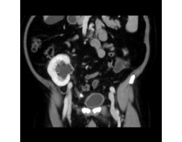
Urolithiasis is a rare urologic complication after kidney transplantation, and its diagnosis and treatment can be challenging for clinicians. In our 52-year-old male patient, graft hydronephrosis was found six months after transplantation. The patient had recurrent urinary tract infections followed by macrohematuria and an increase in creatinine levels. Computerized tomography revealed a 13-mm diameter stone in the ureter of the transplanted kidney as the cause of obstruction. Percutaneous nephrostomy was placed in the graft to solve the obstruction. Initial endoscopic treatment with a retrograde approach failed. An antegrade approach through a previously placed nephrostomy was not successful either. By a repeated retrograde approach, laser lithotripsy was performed successfully. The patient has been monitored for six months and has stable graft function without hydronephrosis or stones. As in our patient's case, the diagnosis and treatment of urolithiasis in kidney transplant patients is challenging, and minimally invasive procedures are the treatment of choice.
Keywords: Kidney transplantation; Laser lithotripsy; Ureterolithiasis; Ureteroscopy.
Sestre Milosrdnice University Hospital.
Figures


Similar articles
-
Minimally invasive procedures for treatment of urolithiasis in transplanted kidneys.Exp Clin Transplant. 2014 Jun;12(3):200-4. Exp Clin Transplant. 2014. PMID: 24907719
-
Over 30-yr Experience on the Management of Graft Stones After Renal Transplantation.Eur Urol Focus. 2018 Mar;4(2):169-174. doi: 10.1016/j.euf.2018.06.007. Epub 2018 Jun 23. Eur Urol Focus. 2018. PMID: 29941388
-
Urolithiasis in Renal Allografts: Complications and Outcomes.Exp Clin Transplant. 2017 Apr;15(2):164-170. doi: 10.6002/ect.2016.0040. Epub 2016 Nov 18. Exp Clin Transplant. 2017. PMID: 27855586
-
Urolithiasis in renal transplant donors and recipients: An update.Int J Surg. 2016 Dec;36(Pt D):693-697. doi: 10.1016/j.ijsu.2016.11.032. Epub 2016 Nov 14. Int J Surg. 2016. PMID: 27856353 Review.
-
Urolithiasis in children: surgical approach.Pediatr Clin North Am. 2012 Aug;59(4):897-908. doi: 10.1016/j.pcl.2012.05.019. Epub 2012 Jun 22. Pediatr Clin North Am. 2012. PMID: 22857836 Review.
References
-
- Orlić L, Jelić Pranjić I. Kidney transplantation in the elderly. Med Flum. 2020;56(4):504–12. [in Croatian]
-
- Rahelić D, Sotošek S, Galić J, Fučkar Ž. Urolitijaza. In: Fučkar Ž, Španjol J, editors. Urologija II. (specijalni dio). 2nd edn. Rijeka: School of Medicine, University of Rijeka, 2013;289-330. (in Croatian)
Publication types
MeSH terms
LinkOut - more resources
Full Text Sources
Medical
Research Materials