Review
. 2024 Jun;19(3):100922.
doi: 10.1016/j.ajps.2024.100922. Epub 2024 Apr 24.

Glucocorticoids-based prodrug design: Current strategies and research progress

Affiliations
Review

Glucocorticoids-based prodrug design: Current strategies and research progress

Hongbing Liu et al. Asian J Pharm Sci. 2024 Jun.

Abstract

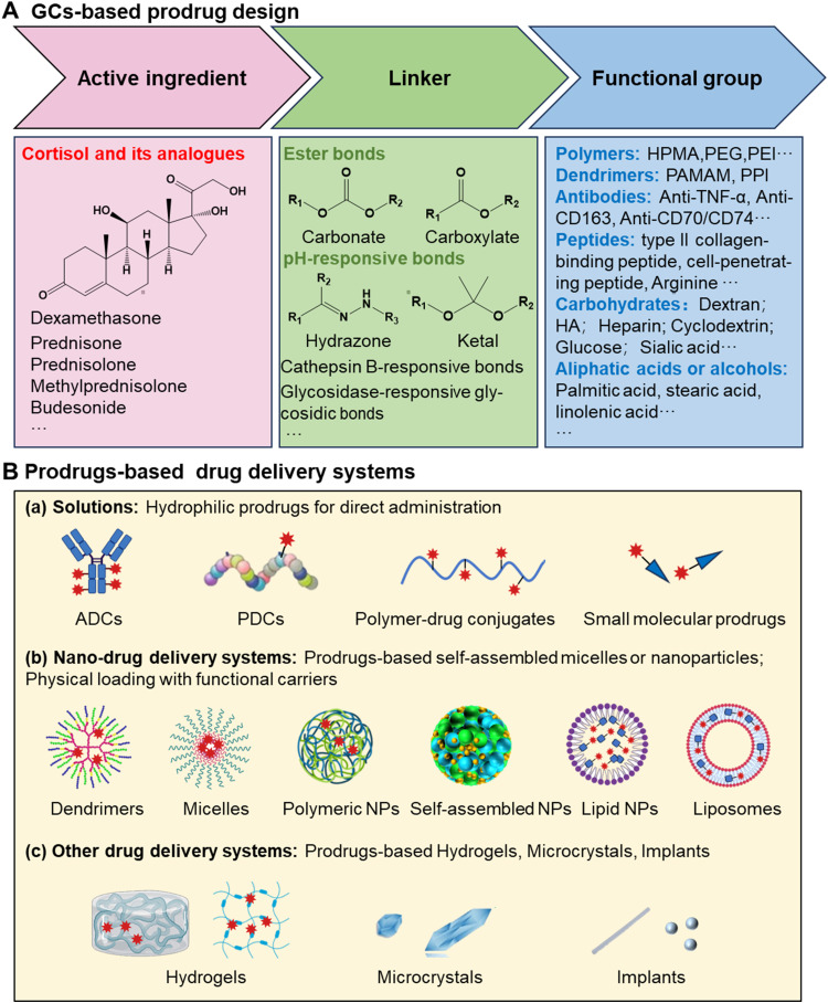
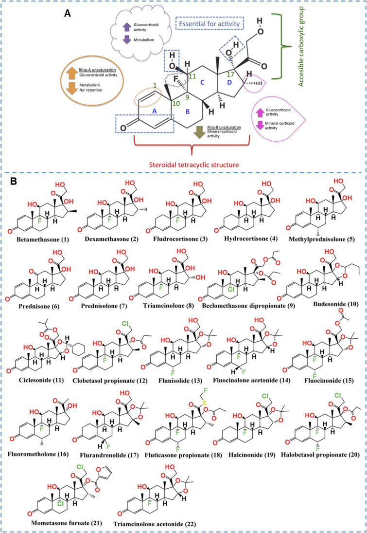
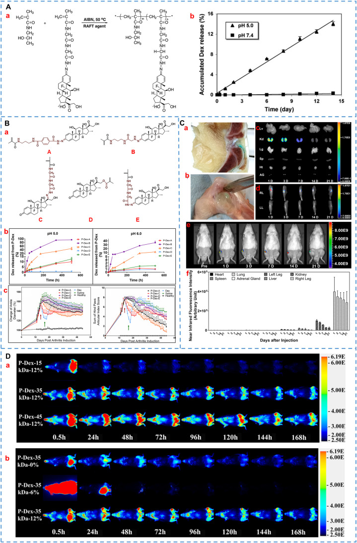
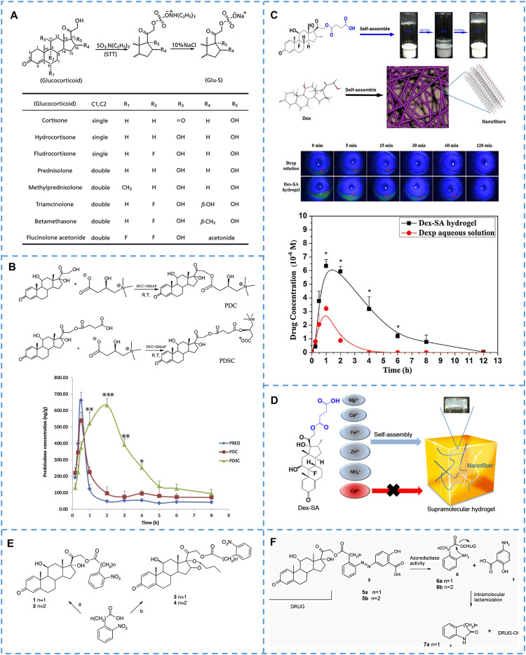
Attributing to their broad pharmacological effects encompassing anti-inflammation, antitoxin, and immunosuppression, glucocorticoids (GCs) are extensively utilized in the clinic for the treatment of diverse diseases such as lupus erythematosus, nephritis, arthritis, ulcerative colitis, asthma, keratitis, macular edema, and leukemia. However, long-term use often causes undesirable side effects, including metabolic disorders-induced Cushing's syndrome (buffalo back, full moon face, hyperglycemia, etc.), osteoporosis, aggravated infection, psychosis, glaucoma, and cataract. These notorious side effects seriously compromise patients' quality of life, especially in patients with chronic diseases. Therefore, glucocorticoid-based advanced drug delivery systems for reducing adverse effects have received extensive attention. Among them, prodrugs have the advantages of low investment, low risk, and high success rate, making them a promising strategy. In this review, we propose the strategies for the design and summarize current research progress of glucocorticoid-based prodrugs in recent decades, including polymer-based prodrugs, dendrimer-based prodrugs, antibody-drug conjugates, peptide-drug conjugates, carbohydrate-based prodrugs, aliphatic acid-based prodrugs and so on. Besides, we also raise issues that need to be focused on during the development of glucocorticoid-based prodrugs. This review is expected to be helpful for the research and development of novel GCs and prodrugs.

Keywords: Glucocorticoids; Prodrug design; Research progress; Targeted drug delivery.


Conflict of interest statement

There are no conflicts to declare.

Figures

Image, graphical abstract
Graphical abstract
Scheme 1
Scheme 1
Schematic illustration of advantages and classification of GCs-based prodrugs.
Fig 1
Fig. 1
(A) Strategies in prodrug design of GCs and (B) prodrug-based drug delivery systems.
Fig 2
Fig. 2
Structure-activity relationship of GCs and structures of the screened FDA-approved GCs. (A) Structure-activity relationship of GCs, Dex as an example. Reprinted with permission , copyright 2016, Elsevier. (B) Chemical structures of the GCs (1∼22) approved by FDA. Reprinted with permission , copyright 2021, The Royal Society of Chemistry.
Fig 3
Fig. 3
Representative HPMA-based GCs prodrugs. (A)(a) Synthesis process of HPMA–Dex conjugates. (b) In vitro release profiles of P-Dex in pH 5.0 and 7.4 buffers. Reprinted with permission , copyright 2008, Springer Nature. (B)(a) Design of Dex-containing monomers (A, B, C, D, E) of HPMA-Dex prodrugs with different releasing rates. (b) Dex release behavior of various P-Dex prodrugs in the different mediums. (c) Pharmacodynamic results of different P-Dex prodrugs on joint inflammation in AIA rats. Reprinted with permission , copyright 2020, Elsevier. (C) Retention of the ProGel-Dex in arthritic joints. (a, b) Retention of ProGel-Dex in the knee joints and dissected ankle joints of rats was observed at 7 d and 28 d after intra-articular injection, respectively. NIR optical images of various organs (c) and hind limbs (d, e) at different time points. (f) The semi-quantitative results of (c) and (d). Reprinted with permission , copyright 2021, Elsevier. (D) The images were acquired following a single intravenous administration of P-Dex-IRDye conjugate with different MW (a) and different Dex content (b). The left and contralateral femurs of mice were challenged with poly (methyl methacrylate) particles and PBS, respectively. Reprinted with permission , copyright 2017, American Chemical Society.
Fig 4
Fig. 4
Representative PEG-based GCs prodrugs. (A) Chemical structure of mPEG-Dex coniugate. Reprinted with permission , copyright 2008, Elsevier. (B) Chemical structure of Click PEG-Dex; The release behaviors of click PEG-Dex at the medium of different pH; Ankle joint diameter changes of the various treatment groups during the entire experiment. Reprinted with permission , copyright 2010, American Chemical Society. (C) Design of ZSJ-0228 that self-assembles into micelles in water, with good renal targeting and the effect of reducing proteinuria caused by nephritis. Reprinted with permission , copyright 2010, American Chemical Society. (D) Chemical structure of PEG-Dex conjugate; pharmacokinetics behaviors and pharmacokinetic parameters after intravenous injection of various Dex-based formulations. Reprinted with permission , copyright 2018, Springer Nature. (E) Chemical structure of SA-PEG-Dex conjugates; the renal fluorescence signal and the semi-quantitative values in AKI mice treated with fluorescent probe-labeled micelles at different time points. Reprinted with permission , copyright 2017, Ivyspring International Publisher.
Fig 5
Fig. 5
Representative polymer-based GCs prodrugs. (A) The synthesis scheme of PEI-Dex. Reprinted with permission , copyright 2007, American Chemical Society. (B) Schematic diagram of preparation and gene delivery of Au-PEI/DNA/PEI-Dex. Reprinted with permission , copyright (2014), American Chemical Society. (C) Synthetic route of Dex-PEI-mannose. Reprinted with permission , copyright 2021, Elsevier. (D) Schematic diagram of prednisolone-PVP prodrug modified nerve electrode. Reprinted with permission , copyright 2010, American Chemical Society. (E) Schematic diagram of preparation and anti-inflammatory treatment of poly-Dex-prodrug nanocapsules. Reprinted with permission , copyright 2021, The Royal Society of Chemistry.
Fig 6
Fig. 6
Representative dendrimer-based GCs prodrugs. (A) Diagrammatic representation of dendrimer-based drug delivery systems. Reprinted with permission , copyright 2021, Elsevier. (B) Synthesis routes of PAMAM G4 (NH2)-Dex conjugate. Reprinted with permission , copyright 2013, Elsevier. (C) Synthetic route of G4(Phe) and G4(Phe)-Dex. Reprinted with permission , copyright 2020, The authors. (D) Preparation of d-Dex and d-Dex-loaded injectable hydrogel. (a, b) Synthesis of d-Dex and gel precursors. (c) Schematic diagram of the formation of a final injectable hydrogel. Reprinted with permission , copyright 2017, Elsevier.
Fig 7
Fig. 7
Representative antibody-drug conjugates of GCs. (A) The chemical structure and mechanism of the anti-CD163- Dex conjugates. Reprinted with permission , copyright 2012, Elsevier. Reprinted with permission , copyright 2016, The authors. (B) Phosphate diester linked α-hCD70 antibody-GCs conjugates. Reprinted with permission , copyright 2016, American Chemical Society. (C) Cathepsin B-cleavable linker and self-immolation spacer linked antibody-budesonide conjugate. Reprinted with permission , copyright 2016, American Chemical Society. (D) Conjugation process for GCs-ADCs; representative structures of budesonide analogues. Reprinted with permission , copyright 2021, American Chemical Society. (E) The structure of anti-α-TNF-Ala-Ala-GCs and therapeutical effects on collagen-induced arthritis mice. Representative pharmacodynamic data of ADCs. Reprinted with permission , copyright 2022, American Chemical Society.
Fig 8
Fig. 8
Representative peptide-based GCs prodrugs. (A) Chemical structure of valine-valine-Dex dipeptide prodrug. Reprinted with permission , copyright 2011, Taylor & Francis. (B) Molecular structure of the Dex-SA-FFFE. Reprinted with permission , copyright 2018, Dove Medical Press Limited. (C) Chemical structure of the cell-penetrating peptide-Dex conjugates. Reprinted with permission , copyright 2020, Elsevier. (D) Schematic representation diagram of l-SS-DEX for modulating the tumor microenvironment. Reprinted with permission , copyright 2020, Elsevier. (E) Diagram of 1-Dex-P self-assembled nanofibers for the treatment of liver fibrosis and therapeutical mechanism. Reprinted with permission , copyright 2018, American Chemical Society. (F) Synthesis of WYRGLRGE-Dex and the interaction between collagen type II. Reprinted with permission , copyright 2019, Elsevier. (G) Diagram of the preparation and targeted treatment for inflammatory vascular diseases of BUD-l-Arg@PSA. Reprinted with permission , copyright 2023, American Chemical Society.
Fig 9
Fig. 9
Representative carbohydrates-based GCs prodrugs. (A) The chemical structure of Dex/prednisolone/Budesonide-β-D-glucoside prodrug. (B) The prodrug structure of budesonide-dextran and the budesonide release in various conditions. Reprinted with permission , copyright 2009, Elsevier. (C) The structure of α-cyclodextrin-succinate-prednisolone prodrug and targeting mechanism after oral administration. Reprinted with permission , copyright 2002, Elsevier. (D) The synthesis and targeting mechanism of Dex-SA. Reprinted with permission , copyright 2022, Elsevier. (E) The preparation of amphiphilic heparin-Dex prodrug and micelles. Reprinted with permission , copyright 2013, Elsevier. (F) The synthesis of prodrug and diagram of the anti-inflammatory effect of MM/HA-Dex. Reprinted with permission , copyright 2022, Elsevier.
Fig 10
Fig. 10
Representative long-chain aliphatic acids or aliphatic alcohols-based GCs prodrugs. (A) DXP-loaded nanoparticles improve pharmacokinetic properties. Reprinted with permission , copyright 2019, American Chemical Society. (B) Preparation and antitumor study of Dex-ALA/DTX nanoparticles. Reprinted with permission , copyright 2022, Elsevier. (C) Structures of lipophilic Dex prodrugs . Reprinted with permission, copyright 2018, Elsevier. (D) Preparation and structural screening of AKP-Dex prodrug nanoparticles. a) AKP-Dexs were synthesized from Dex and isopropenyl ethers and were then co-assembled with DSPE−mPEG2000 by a nanoprecipitation method to form AKP-Dex-loaded NPs. (b) Promoiety structures and designations for the corresponding AKP-Dexs. Screening of promoieties for AKP-Dex-loaded NPs on the basis of size, colloidal stability, and acid sensitivity of the linker. Reprinted with permission , copyright 2020, American Chemical Society. (E) SKD nanocrystals were used to treat arthritis: (a) Preparation of SKD MCs: the larger MCs (3.1 µm) were formulated by anti-solvent crystallization method, and the smaller ones (1.1 µm) were further fabricated by wet grinding of larger MCs. (b) In a CIA rat model, MCs exhibited sustained release of native Dex, biocompatible stearyl alcohol and the metabolite acetone in the acidic inflammatory joints post IA injection. Attributing to the stronger sustained-release effect, the therapeutic effect of MCs with an average particle size of 3.1 µm was better than that of 1.1 µm. Reprinted with permission , copyright 2021, Elsevier.
Fig 11
Fig. 11
Representative Sulfate sodium/L-Carnitine/Succinic acid/Nitrophenyl-based GCs prodrugs. (A) Reaction scheme of sulfate sodium-based GCs prodrugs. Reprinted with permission , copyright 2011, Oxford University Press. (B) The synthesis of PDC and PDSC and mean lung prednisolone concentration-time curves of PRED, PDC, and PDSC solutions. Reprinted with permission , copyright 2011, American Chemical Society. Reprinted with permission , copyright 2014, Elsevier. (C) Preparation, precorneal retention and in vitro drug release of Dex-SA hydrogel. Reprinted with permission , copyright 2018, Elsevier. (D) Dex-SA can form supermolecule hydrogels with various cations. Reprinted with permission , copyright 2018, Elsevier. (E) The chemical structure of the nitro-substituted 21-ester for the GCs. Reprinted with permission , copyright 2013, Elsevier. (F) Cyclization-activated steroid prodrugs for the colon. Reprinted with permission , copyright 2009, American Chemical Society.
Fig 12
Fig. 12
Representative other types of GCs prodrugs. (A) Schematic structure of the dimer prodrug and release mechanism of drug from implants. Reprinted with permission , copyright (2021), Springer Nature. (B) Representative pharmacodynamic results and diagram of the synergistic mechanism of Dex-MMF prodrug. Reprinted with permission , copyright (2021), American Chemical Society. (C) Diagram of the esterase-responsive CD-Dex prodrugs for the treatment of liver fibrosis. Reprinted with permission , copyright (2021), Wiley-VCH GmbH.

References

    1. Rodríguez Villanueva J., Rodríguez Villanueva L., Guzmán Navarro M. Pharmaceutical technology can turn a traditional drug, dexamethasone into a first-line ocular medicine. A global perspective and future trends. Int J Pharm. 2017;516(1–2):342–351. - PubMed
    1. Fung A.T., Tran T., Lim L.L., Samarawickrama C., Arnold J., Gillies M., et al. Local delivery of corticosteroids in clinical ophthalmology: a review. Clin Experiment Ophthalmol. 2020;48(3):366–401. - PMC - PubMed
    1. Meduri G.U., Annane D., Confalonieri M., Chrousos G.P., Rochwerg B., Busby A., et al. Pharmacological principles guiding prolonged glucocorticoid treatment in ARDS. Intensive Care Med. 2020;46(12):2284–2296. - PMC - PubMed
    1. Hardy R.S., Raza K., Cooper M.S. Therapeutic glucocorticoids: mechanisms of actions in rheumatic diseases. Nat Rev Rheumatol. 2020;16(3):133–144. - PubMed
    1. Talebian S., Wallace G.G., Schroeder A., Stellacci F., Conde J. Nanotechnology-based disinfectants and sensors for SARS-CoV-2. Nat Nanotechnol. 2020;15(8):618–621. - PubMed

LinkOut - more resources