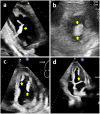
Hypereosinophilia and Left Ventricular Thrombus: A Case Report and Literature Review
- PMID: 38966441
- PMCID: PMC11223752
- DOI: 10.7759/cureus.61674
Hypereosinophilia and Left Ventricular Thrombus: A Case Report and Literature Review
Abstract
Left ventricular thrombus (LVT) has historically been reported as a complication of acute left ventricular (LV) myocardial infarction. It is most commonly observed in cases of LV systolic dysfunction attributed to ischemic or nonischemic etiologies. Conversely, the occurrence of LVT in normal LV systolic function is an exceptionally rare presentation and is predominantly associated with conditions such as hypereosinophilic syndrome (HES), cardiac amyloidosis, left ventricular noncompaction, hypertrophic cardiomyopathy (HCM), hypercoagulability states, immune-mediated disorders, and malignancies. Notably, hypereosinophilia (HE) has been linked with thrombotic events. Intracardiac thrombus is a well-known complication of eosinophilic myocarditis (EM) or Loeffler endomyocarditis, both of which are considered clinical manifestations of HES. We present a case of a 63-year-old male with normal LV systolic function, HE, and noncontributory hypercoagulability workup, who presented with thromboembolic complications arising from LVT. Interestingly, the diagnostic evaluation for EM and Loeffler endocarditis was nonconfirmatory. Additionally, we performed a literature review to delineate all similar cases. This article also outlines the pathophysiology, diagnosis, and treatment approaches for hypereosinophilic cardiac involvement with a specific focus on LVT.
Keywords: absolute eosinophil count; arterial thromboembolism; cardiac magnetic resonance imaging (cmri); endomyocardial biopsy; eosinophilic myocarditis; hypereosinophilia; hypereosinophilia syndrome; left ventricular thrombosis; loeffler endomyocarditis; restrictive cardiomyopathy.
Copyright © 2024, Khachatryan et al.
Conflict of interest statement
Human subjects: Consent was obtained or waived by all participants in this study. Conflicts of interest: In compliance with the ICMJE uniform disclosure form, all authors declare the following: Payment/services info: All authors have declared that no financial support was received from any organization for the submitted work. Financial relationships: All authors have declared that they have no financial relationships at present or within the previous three years with any organizations that might have an interest in the submitted work. Other relationships: All authors have declared that there are no other relationships or activities that could appear to have influenced the submitted work.
Figures


Similar articles
-
The Cleveland Clinic experience of eosinophilic myocarditis in the setting of hypereosinophilic syndrome: demographics, cardiac imaging, and outcomes.Cardiovasc Diagn Ther. 2024 Dec 31;14(6):1122-1133. doi: 10.21037/cdt-24-347. Epub 2024 Dec 2. Cardiovasc Diagn Ther. 2024. PMID: 39790190 Free PMC article.
-
Normalization of left ventricular filling pressure after cardiac surgery for the Loeffler's endocarditis: a case report.Eur Heart J Case Rep. 2021 Jun 28;5(6):ytab189. doi: 10.1093/ehjcr/ytab189. eCollection 2021 Jun. Eur Heart J Case Rep. 2021. PMID: 34263118 Free PMC article.
-
Loeffler endocarditis with intracardiac thrombus: case report and literature review.BMC Cardiovasc Disord. 2021 Dec 28;21(1):615. doi: 10.1186/s12872-021-02443-2. BMC Cardiovasc Disord. 2021. PMID: 34961478 Free PMC article.
-
Emerging Trends in Left Ventricular Thrombus: A Comprehensive Review of Non-Ischemic and Ischemic Cardiopathies, Including Eosinophilic Myocarditis, Chagas Cardiomyopathy, Amyloidosis, and Innovative Anticoagulant Approaches.Diagnostics (Basel). 2024 Apr 30;14(9):948. doi: 10.3390/diagnostics14090948. Diagnostics (Basel). 2024. PMID: 38732361 Free PMC article. Review.
-
Loeffler endocarditis as a rare cause of heart failure with preserved ejection fraction: A case report and review of literature.Medicine (Baltimore). 2018 Mar;97(11):e0079. doi: 10.1097/MD.0000000000010079. Medicine (Baltimore). 2018. PMID: 29538200 Free PMC article.
Cited by
-
Multimodality Imaging in Eosinophilic Myocarditis: A Rare Cause of Heart Failure.J Cardiovasc Dev Dis. 2025 Aug 21;12(8):320. doi: 10.3390/jcdd12080320. J Cardiovasc Dev Dis. 2025. PMID: 40863386 Free PMC article. Review.
References
-
- World Health Organization-defined eosinophilic disorders: 2022 update on diagnosis, risk stratification, and management. Shomali W, Gotlib J. Am J Hematol. 2022;97:129–148. - PubMed
-
- Hypereosinophilic syndrome and thrombosis: a retrospective review. Wallace KL, Elias MK, Butterfield JH, Weiler CR. https://doi.org/10.1016/j.jaci.2012.12.1105 J Allergy Clin Immunol. 2013;131:0.
Publication types
LinkOut - more resources
Full Text Sources
