

High-frequency ultrasound accuracy in preoperative cutaneous melanoma assessment: A meta-analysis
- PMID: 38967397
- PMCID: PMC11664467
- DOI: 10.1111/jdv.20179
High-frequency ultrasound accuracy in preoperative cutaneous melanoma assessment: A meta-analysis
Abstract
Background: High-frequency ultrasound (HFUS) can safely and efficiently visualize cutaneous tumour characteristics including depth.
Objectives: We aimed to evaluate its accuracy in measuring melanoma depth against the gold standard, histopathology, for treatment planning.
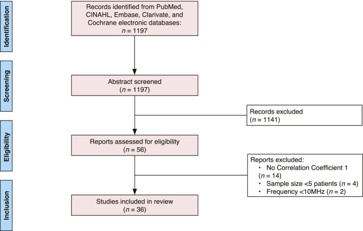
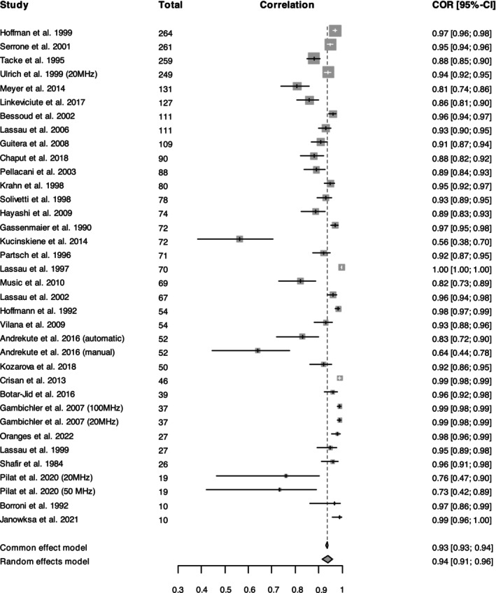
Methods: A review of publications was conducted in March 2023 through five electronic databases. Thirty-six included articles studied patients who received HFUS (≥10 MHz) measurements, melanoma biopsy or excision, and reported a tumour depth correlation coefficient between HFUS and histopathology. We analysed correlation coefficients between HFUS and histopathology, measured tumour depths and shed light on reasons for mismeasurements. Additionally, we identified the reporting of critical metrics including, lesion characteristics, melanoma subtype, type of correlation coefficient, 95% confidence intervals for Pearson coefficients and sample size.
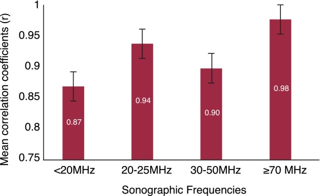
Results: The most common tumour imaged was superficial spreading melanoma on the trunk and extremities, followed by head/face. Maximum ultrasound frequencies ranged from 13 MHz to 100 MHz with participants ranging from 5 to 264. Histopathology and HFUS correlation coefficients ranged from 0.417 to 0.997 (median: 0.94, mean: 0.89 and SD: 0.13). Lower frequency probes (10-20 MHz) were less accurate in assessing melanoma thickness, with a cumulative mean correlation coefficient of 0.87 compared to 0.94 (20-25 MHz) and 0.98 (≥70 MHz). Studies demonstrated higher sonographic accuracy in melanomas >0.75 mm. Additionally, ultrasound may report increased melanoma depth compared to histopathology for reasons including lymphocytic infiltration, presence of a nevus and shrinkage during specimen processing. Furthermore, we found a gap in the reporting of details such as fundamental characteristics of lesion populations. Specifically, 86% (31 out of 36) of the studies failed to report one or more critical metrics, such as mean, median or range of lesion depths.
Conclusions: HFUS may serve as a supplementary tool for preoperative melanoma assessment, with increased accuracy in thicker tumours. Frequencies <20 MHz are less reliable in assessing depth. Frequencies ≥70 MHz demonstrate stronger correlations to histopathology. Higher ultrasound accuracy was seen for melanomas with Breslow depth >0.75 mm.
© 2024 The Author(s). Journal of the European Academy of Dermatology and Venereology published by John Wiley & Sons Ltd on behalf of European Academy of Dermatology and Venereology. This article has been contributed to by U.S. Government employees and their work is in the public domain in the USA.
Conflict of interest statement
The authors declare no conflict of interest.
Figures

Similar articles
-
High-frequency ultrasound for diagnosing skin cancer in adults.Cochrane Database Syst Rev. 2018 Dec 4;12(12):CD013188. doi: 10.1002/14651858.CD013188. Cochrane Database Syst Rev. 2018. PMID: 30521683 Free PMC article.
-
Assessment of tumor thickness in melanocytic skin lesions: comparison of optical coherence tomography, 20-MHz ultrasound and histopathology.Dermatology. 2011;223(2):161-8. doi: 10.1159/000332845. Epub 2011 Oct 20. Dermatology. 2011. PMID: 22024981
-
A Preliminary Study for Quantitative Assessment with HFUS (High- Frequency Ultrasound) of Nodular Skin Melanoma Breslow Thickness in Adults Before Surgery: Interdisciplinary Team Experience.Curr Radiopharm. 2020;13(1):48-55. doi: 10.2174/1874471012666191007121626. Curr Radiopharm. 2020. PMID: 31589132
-
Preoperative 15-MHz ultrasound assessment of tumor thickness in malignant melanoma.Actas Dermosifiliogr. 2013 Apr;104(3):227-31. doi: 10.1016/j.ad.2012.06.007. Epub 2012 Aug 28. Actas Dermosifiliogr. 2013. PMID: 22938997 English, Spanish.
-
Role of In Vivo Reflectance Confocal Microscopy in the Analysis of Melanocytic Lesions.Acta Dermatovenerol Croat. 2018 Apr;26(1):64-67. Acta Dermatovenerol Croat. 2018. PMID: 29782304 Review.
Cited by
-
Advances in Skin Ultrasonography for Malignant and Benign Tumors of the Head and Neck: Current Insights and Future Directions.J Clin Med. 2025 Mar 27;14(7):2298. doi: 10.3390/jcm14072298. J Clin Med. 2025. PMID: 40217748 Free PMC article. Review.
-
Ultrasound in Skin Cancer: Why, How, and When to Use It?Cancers (Basel). 2024 Sep 27;16(19):3301. doi: 10.3390/cancers16193301. Cancers (Basel). 2024. PMID: 39409920 Free PMC article. Review.
References
-
- Thompson JF, Soong SJ, Balch CM, Gershenwald JE, Ding S, Coit DG, et al. Prognostic significance of mitotic rate in localized primary cutaneous melanoma: an analysis of patients in the multi‐institutional American joint committee on cancer melanoma staging database. JCO. 2011;29(16):2199–2205. - PMC - PubMed
-
- Ward WH, Farma JM, editors. Cutaneous melanoma: etiology and therapy. Philadelphia, USA: Codon Publications; 2017. [cited 2023 Aug 25]. Available from: https://exonpublications.com/index.php/exon/issue/view/8 - PubMed
Publication types
MeSH terms
Grants and funding
LinkOut - more resources
Full Text Sources
Medical
Miscellaneous
