. 2024 Oct;67(10):2260-2274.
doi: 10.1007/s00125-024-06207-3. Epub 2024 Jul 5.

tRNA-derived fragments in T lymphocyte-beta cell crosstalk and in type 1 diabetes pathogenesis in NOD mice

Affiliations

tRNA-derived fragments in T lymphocyte-beta cell crosstalk and in type 1 diabetes pathogenesis in NOD mice

Flora Brozzi et al. Diabetologia. 2024 Oct.

Abstract

Aims/hypothesis: tRNAs play a central role in protein synthesis. Besides this canonical function, they were recently found to generate non-coding RNA fragments (tRFs) regulating different cellular activities. The aim of this study was to assess the involvement of tRFs in the crosstalk between immune cells and beta cells and to investigate their contribution to the development of type 1 diabetes.

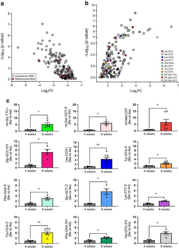
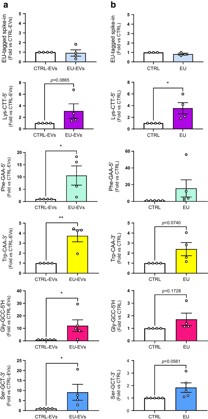
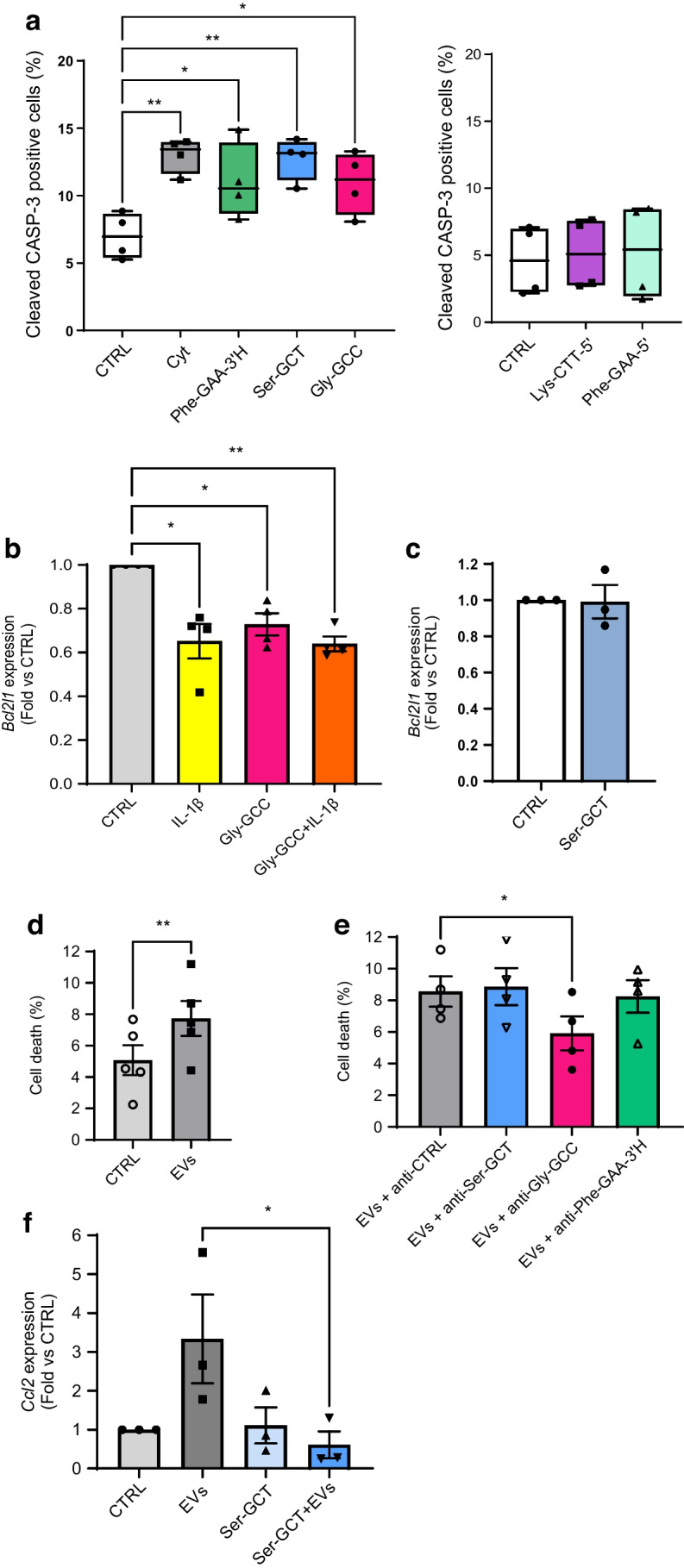
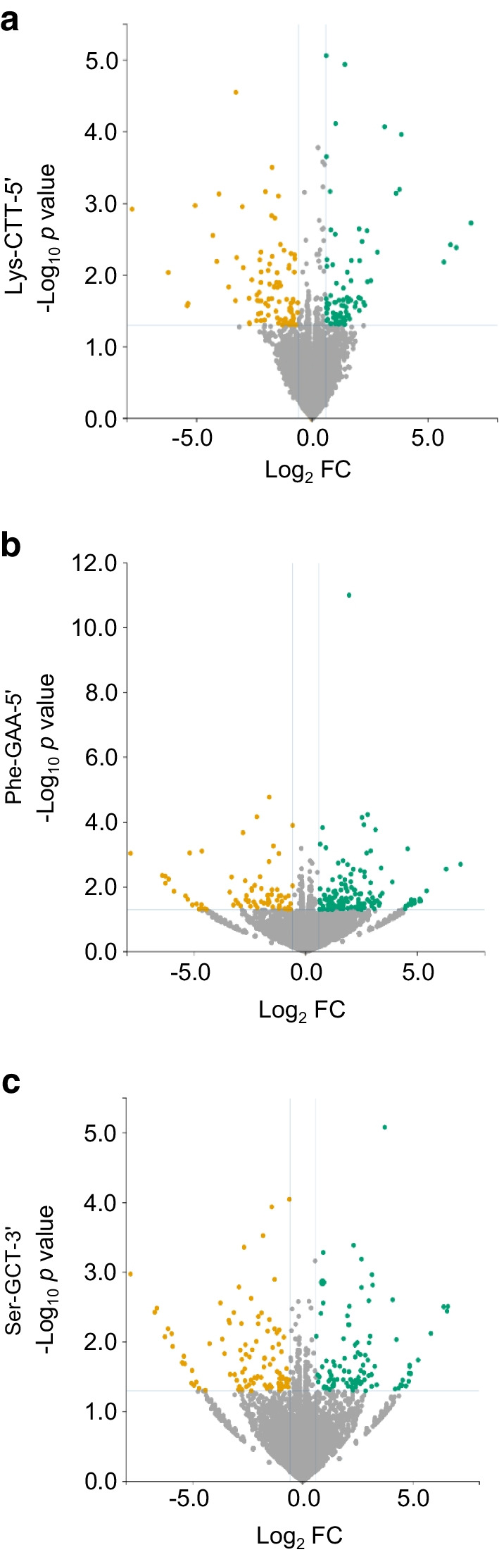
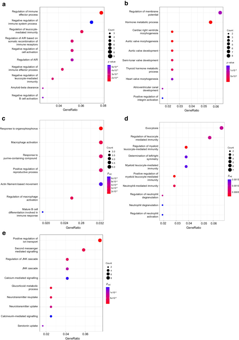
Methods: Global profiling of the tRFs present in pancreatic islets of 4- and 8-week-old NOD mice and in extracellular vesicles released by activated CD4+ T lymphocytes was performed by small RNA-seq. Changes in the level of specific fragments were confirmed by quantitative PCR. The transfer of tRFs from immune cells to beta cells occurring during insulitis was assessed using an RNA-tagging approach. The functional role of tRFs increasing in beta cells during the initial phases of type 1 diabetes was determined by overexpressing them in dissociated islet cells and by determining the impact on gene expression and beta cell apoptosis.

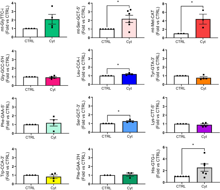
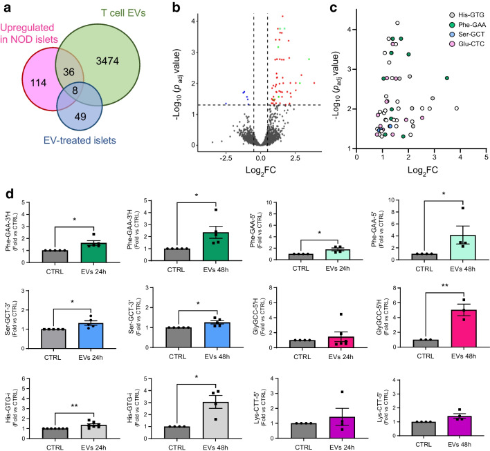
Results: We found that the tRF pool was altered in the islets of NOD mice during the initial phases of type 1 diabetes. Part of these changes were triggered by prolonged exposure of beta cells to proinflammatory cytokines (IL-1β, TNF-α and IFN-γ) while others resulted from the delivery of tRFs produced by CD4+ T lymphocytes infiltrating the islets. Indeed, we identified several tRFs that were enriched in extracellular vesicles from CD4+/CD25- T cells and were transferred to beta cells upon adoptive transfer of these immune cells in NOD.SCID mice. The tRFs delivered to beta cells during the autoimmune reaction triggered gene expression changes that affected the immune regulatory capacity of insulin-secreting cells and rendered the cells more prone to apoptosis.

Conclusions/interpretation: Our data point to tRFs as novel players in the crosstalk between the immune system and insulin-secreting cells and suggest a potential involvement of this novel class of non-coding RNAs in type 1 diabetes pathogenesis.

Data availability: Sequences are available from the Gene Expression Omnibus (GEO) with accession numbers GSE242568 and GSE256343.

Keywords: Apoptosis; Autoimmunity; Extracellular vesicles; Insulin; Pancreatic islet.


Figures

Fig. 1
Fig. 1
Identification of tRFs displaying changes in their level in the islets of prediabetic NOD mice. (a) tRFs significantly downregulated in the islets of 8-week-old mice compared with the islets of 4-week-old mice. (b) tRFs significantly upregulated in islets of 8-week-old mice compared with the islets of 4-week-old mice. Squares, tRFs derived from mitochondrially encoded tRNAs; circles, cytoplasmic tRFs. Colour codes indicate fragments originating from the same isoacceptor tRNA. (c) Real-time PCR confirmation of the upregulation of selected tRFs in islets of 8-week-old mice. The data were normalised to the level of the small ncRNA miR-184–5p, which displays no changes under prediabetic conditions. The level of the fragments in 4-week-old mice has been set to 1 and the data shown are the means ± SD. n=3–7. *p<0.05, **p<0.01 (unpaired t test). FC, fold change
Fig. 2
Fig. 2
Identification of tRFs displaying changes in their levels in cytokine-treated islet cells. C57BL/6NRj mouse islet cells incubated with or without a mix of cytokines (IL-1β, IFN-γ and TNF-α) for 24 h before RNA collection. Real-time PCR validation of tRFs differentially expressed in cytokine-treated islet cells (Cyt) compared with control (CTRL). The data were normalised to the level of miR-7a and miR-375, which displays no changes in response to proinflammatory cytokines. The data are shown as fold changes vs CTRL and are presented as mean ± SD. n=3–6. *p<0.05 (paired t test). FC, fold change
Fig. 3
Fig. 3
T cell EVs contain tRFs that are transferred to islet cells resulting in changes in the tRF pool. (a) Venn diagram showing the number of tRFs present in T cell EVs displaying changes in the islets of prediabetic NOD mice and in EV-treated islet cells. (b) Volcano plot indicating the changes in tRF level in islet cells after 24 h incubation with NOD mouse T cell EVs. Fragments above the horizontal dashed line display significant changes (padj<0.05). Vertical dashed lines indicate a fold change of ±2. (c) Significantly upregulated tRFs in EV-treated mouse islet cells. Selected tRFs that originate from the same isodecoder tRNA are labelled with the same colour. (d) Analysis by qPCR of the level of selected tRFs in islet cells after 24 h and 48 h incubation with EVs released by NOD mouse T cells. The data were normalised to the level of miR-7a and miR-375, which display no changes under these experimental conditions; values are shown as fold change vs CTRL and are presented as mean ± SD. n=3 or 6. *p<0.05, **p<0.01 (ratio paired t test). FC, fold change
Fig. 4
Fig. 4
tRFs of CD4+/CD25 T cells transferred in vitro and in vivo to pancreatic beta cells. (a) Real-time PCR of islet cell tRFs pulled down on streptavidin beads upon incubation with EVs released by EU-treated T cells (EU-EVs) or untreated control T cells (CTRL-EVs). An EU-tagged C. elegans miR-238 mimic was spiked in cell extracts as internal control. *p<0.05, **p<0.01 (ratio paired t test). (b) EU-tagged RNAs from FAC-sorted beta cells of NOD.SCID mice injected with EU-tagged T cells (EU) or with saline solution (CTRL) were purified on streptavidin beads and analysed by qPCR. An EU-tagged oligonucleotide containing the sequence of C. elegans miR-238 was spiked in the samples and was used as internal control to normalise the data. Data are presented as mean ± SD. n=3–5. *p<0.05 (ratio paired t test)
Fig. 5
Fig. 5
Modulation of the levels of selected tRFs affects beta cell apoptosis. (a) Mouse islet cells were transfected with different tRF mimics (as indicated) or with a scrambled control sequence (CTRL) for 48 h. Part of the cells were incubated for 24 h with a mix of proinflammatory cytokines (IL-1β, IFN-γ and TNF-α) (Cyt) prior to staining using antibodies against insulin and cleaved caspase-3 (CASP-3). Between 600 and 1000 cells per condition were counted and the percentage of cleaved caspase-3 positive beta cells was calculated. *p<0.05, **p<0.01 (one-way ANOVA), n=4 (Dunnett Correction). (b) MIN6 cells were transfected with an oligonucleotide containing the sequence of Gly-GCC-5′H (Gly-GCC) or with a scrambled sequence (CTRL) for 48 h and were subsequently incubated with or without IL-1β for 24 h. RNA was collected and Bcl2l1 expression was measured by qPCR. *p<0.05, **p<0.01 (one-way ANOVA), n=4 (Dunnett correction). (c) MIN6 cells were transfected with an oligonucleotide containing the sequence of Ser-GCT or with a scrambled sequence (CTRL). Two days later, RNA was collected and Bcl2l1 expression was measured by qPCR. (d, e) Mouse pancreatic islet cells were transfected with a control oligonucleotide inhibitor (CTRL) or with a tRF inhibitor as indicated. After 24 h, the cells were treated with T cell EVs and incubated for another 48 h. Islet cell death was assessed by scoring the cells displaying pycnotic nuclei upon Hoechst/propidium iodide staining (around 5000 cells were counted per condition). **p<0.01 (paired t test), n=5 (d); *p<0.05 (one-way ANOVA, Dunnett post hoc test), n=4 (e). (f) MIN6 cells were transfected with Ser-GCT-3′ inhibitor or a negative control (CTRL). After 24 h, the cells were incubated with or without T cell EVs for another 24 h. RNA was collected and Ccl2 expression was measured by qPCR. *p<0.05 (one-way ANOVA, Šidák correction), n=3. (a) Data are presented as median, with 25th and 75th percentile, and whiskers showing minimum and maximum. (bf) Data are presented as mean ± SD
Fig. 6
Fig. 6
Overexpression of Lys-CTT-5′, Phe-GAA-5′ or Ser-GCT-3′ affects islet cell gene expression. Volcano plots of the differentially expressed transcripts in Lys-CTT-5′ (a), Phe-GAA-5′ (b) or Ser-GCT-3′ (c) overexpressing islet cells compared with cells transfected with a scrambled oligonucleotide. Upregulated transcripts are shown in green and downregulated transcripts are shown in orange. FC, fold change
Fig. 7
Fig. 7
GO analysis of the transcripts affected by the overexpression of Lys-CTT-5′, Phe-GAA-5′ or Ser-GCT-3′. (a, b) Top enriched GO terms for downregulated (a) and upregulated (b) genes in mouse islet cells overexpressing Lys-CTT-5′. (c, d) Top enriched GO terms for downregulated (c) and upregulated genes (d) in mouse islet cells overexpressing Phe-GAA-5′. (e) Top enriched GO terms for upregulated genes in mouse islet cells overexpressing Ser-GCT-3′. The size of the circles is proportional to the ratio of the observed vs the expected overlaps. p values correspond to hypergeometric tests (colour-coded)

References

    1. Quattrin T, Mastrandrea LD, Walker LSK (2023) Type 1 diabetes. Lancet 401(10394):2149–2162. 10.1016/S0140-6736(23)00223-4 - PubMed
    1. van Belle TL, Coppieters KT, von Herrath MG (2011) Type 1 diabetes: etiology, immunology, and therapeutic strategies. Physiol Rev 91(1):79–118. 10.1152/physrev.00003.2010 - PubMed
    1. Eizirik DL, Colli ML, Ortis F (2009) The role of inflammation in insulitis and beta-cell loss in type 1 diabetes. Nat Rev Endocrinol 5(4):219–226. 10.1038/nrendo.2009.21 - PubMed
    1. Guay C, Kruit JK, Rome S et al (2019) Lymphocyte-derived exosomal microRNAs promote pancreatic beta cell death and may contribute to type 1 diabetes development. Cell Metab 29(2):348–361, e346. 10.1016/j.cmet.2018.09.011 - PubMed
    1. Gonzalez-Moro I, Olazagoitia-Garmendia A, Colli ML et al (2020) The T1D-associated lncRNA Lnc13 modulates human pancreatic beta cell inflammation by allele-specific stabilization of STAT1 mRNA. Proc Natl Acad Sci U S A 117(16):9022–9031. 10.1073/pnas.1914353117 - PMC - PubMed

MeSH terms