

Urgent and emergent pediatric cardiovascular imaging
- PMID: 38967787
- PMCID: PMC11982110
- DOI: 10.1007/s00247-024-05980-y
Urgent and emergent pediatric cardiovascular imaging
Abstract
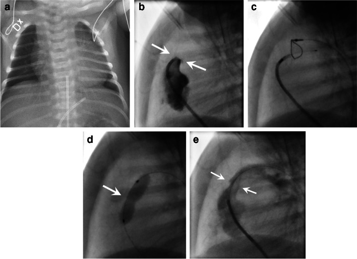
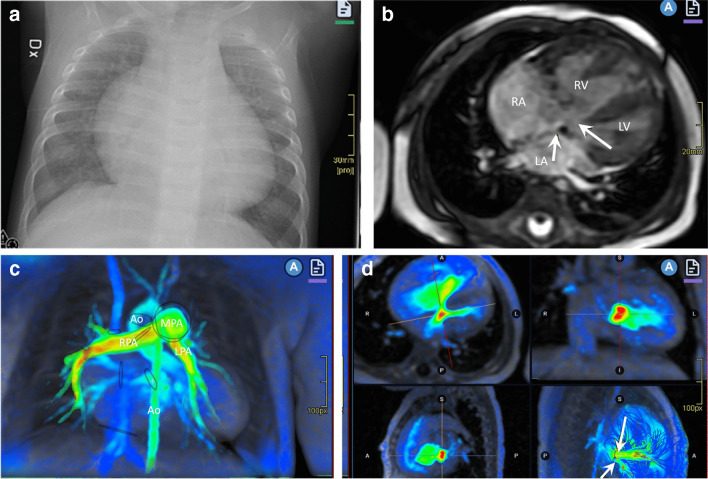
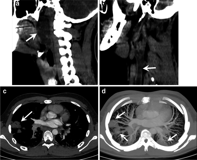
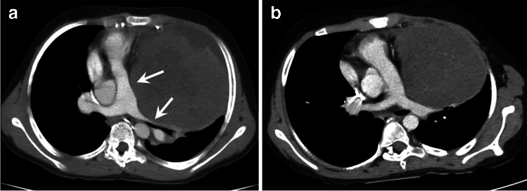
The need for urgent or emergent cardiovascular imaging in children is rare when compared to adults. Patients may present from the neonatal period up to adolescence, and may require imaging for both traumatic and non-traumatic causes. In children, coronary pathology is rarely the cause of an emergency unlike in adults where it is the main cause. Radiology, including chest radiography and computed tomography in conjunction with echocardiography, often plays the most important role in the acute management of these patients. Magnetic resonance imaging can occasionally be useful and may be suitable in more subacute cases. Radiologists' knowledge of how to manage and interpret these acute conditions including knowing which imaging technique to use is fundamental to appropriate care. In this review, we will concentrate on the most common cardiovascular emergencies in the thoracic region, including thoracic traumatic and non-traumatic emergencies and pulmonary vascular emergencies, as well as acute clinical disorders as a consequence of primary and postoperative congenital heart disease. This review will cover situations where cardiovascular imaging may be acutely needed, and not strictly emergencies only. Imaging recommendations will be discussed according to the different clinical presentations and underlying pathology.
Keywords: Cardiovascular system; Computed tomography; Congenital heart defect; Emergencies; Imaging; Pediatric.
© 2024. The Author(s).
Conflict of interest statement
Declarations. Conflicts of interest: None
Figures


References
-
- Han BK, Rigsby CK, Hlavacek A et al (2015) Computed tomography imaging in patients with congenital heart disease part I: rationale and utility. An expert consensus document of the Society of Cardiovascular Computed Tomography (SCCT): Endorsed by the Society of Pediatric Radiology (SPR) and the North American Society of Cardiac Imaging (NASCI). J Cardiovasc Comput Tomogr 9:475–492 - PubMed
-
- Tivnan P, Winant AJ, Johnston PR et al (2021) Thoracic CTA in infants and young children: image quality of dual-source CT (DSCT) with high-pitch spiral scan mode (turbo flash spiral mode) with or without general anesthesia with free-breathing technique. Pediatr Pulmonol 56:2660–2667 - PubMed
-
- Kino A, Zucker EJ, Honkanen A et al (2019) Ultrafast pediatric chest computed tomography: comparison of free-breathing vs. breath-hold imaging with and without anesthesia in young children. Pediatr Radiol 49:301–307 - PubMed
-
- Goo HW (2018) Image quality and radiation dose of high-pitch dual-source spiral cardiothoracic computed tomography in young children with congenital heart disease: comparison of non-electrocardiography synchronization and prospective electrocardiography triggering. Korean J Radiol 19:1031–1041 - PMC - PubMed
Publication types
MeSH terms
LinkOut - more resources
Full Text Sources
Medical
