. 2024;11(5):935-957.
doi: 10.3233/JND-230021.

Phenotype-Genotype Correlation of a Cohort of Patients with Congenital Myopathy: A Single Centre Experience from India

Affiliations

Phenotype-Genotype Correlation of a Cohort of Patients with Congenital Myopathy: A Single Centre Experience from India

Ganaraja Valakunja Harikrishna et al. J Neuromuscul Dis. 2024.

Abstract

Background: Congenital myopathies (CMs) are a diverse group of inherited muscle disorders with broad genotypic and phenotypic heterogeneity. While the literature on CM is available from European countries, comprehensive data from the Indian subcontinent is lacking.

Objectives: This study aims to describe the clinical and histopathological characteristics of a cohort of genetically confirmed CMs from India and attempts to do phenotype-genotype correlation.

Methods: A retrospective chart review of genetically confirmed CMs was evaluated between January 2016 and December 2020 at the neuromuscular clinic. The clinical, genetic, and follow-up data were recorded in a pre-structured proforma as per the medical records, and the data was analyzed.

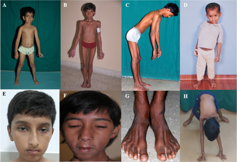
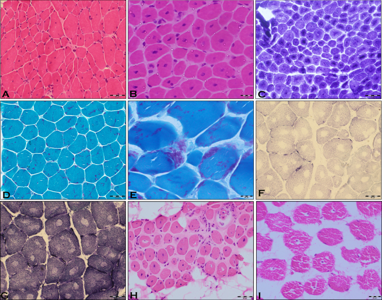
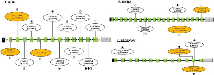
Results: A total of 31(M: F = 14 : 17) unrelated patients were included. The median age at onset and duration of illness are 2.0(IQR:1-8) years and 6.0(IQR:3-10) years respectively. Clinical features observed were proximodistal weakness (54.8%), facial weakness (64.5%), and myopathic facies (54.8%), followed by ptosis (33.3%), and ophthalmoplegia (19.4%). Muscle histopathology was available in 38.7% of patients, and centronuclear myopathy was the most common histopathology finding. The pathogenic genetic variants were identified in RYR1 (29.0%), DNM2 (19.4%), SELENON (12.9%), KBTBD13 (9.7%), NEB (6.5%), and MYPN (6.5%) genes. Novel mutations were observed in 30.3% of the cohort. Follow-up details were available in 77.4% of children, and the median duration of follow-up and age at last follow-up was 4.5 (Range 0.5-11) years and 13 (Range 3-35) years, respectively. The majority were ambulant with minimal assistance at the last follow-up. Mortality was noted in 8.3% due to respiratory failure in Centronuclear myopathy 1 and congenital myopathy 3 with rigid spines (SELENON).

Conclusion: This study highlights the various phenotypes and patterns of genetic mutations in a cohort of pediatric patients with congenital myopathy from India. Centronuclear myopathy was the most common histological classification and the mutations in RYR1 followed by DNM2 gene were the common pathogenic variants identified. The majority were independent in their activities of daily living during the last follow-up, highlighting the fact that the disease has slow progression irrespective of the genotype.

Keywords: Congenital myopathy; DMN2 gene; KBTBD13 gene; RYR1 gene; SELENON gene; creatine phosphokinase; histopathology; phenotype-genotype.


Conflict of interest statement

Nil.

Figures

Fig. 1
Fig. 1
Representative photographs of children with congenital myopathy showing features of – A. dental mal-occlusion and bilateral ptosis (P-14), B: myopathic facies as elongated face and sunken cheeks (P-18), C. rigid spine (P-19), D. skeletal deformity- scoliosis (P-8), E bilateral ptosis (P-28), F. bifacial weakness(P-18), G. wasting of extensor digitorum brevis in both legs (P-19), H. features suggesting proximal lower limb weakness on trying to get up from floor (P-20). (Consent obtained)
Fig. 2
Fig. 2
Microphotograph showing transverse section of P-2, centronuclear myopathy with many muscle fibres (>30%) possessing central nuclei (H & E x100) [A, B] and radial staining pattern (NADH × 200) [C]. Microphotograph showing transverse section in P-25, of Nemaline myopathy with many muscle fibres displaying Nemaline rods of variable density, configuration and distribution (MGT × 200) [D]displaying Nemaline rods (black arrow) of variable density, configuration and distribution (MGT × 400) [E]. Microphotograph showing transverse section of P-27, central core myopathy with many muscle fibres displaying central areas (Cores) of absence of oxidative activity (SDH × 200) [F] and (NADH × 200) [G]. Note the type I fibre predominance. Microphotograph showing transverse section in P-15 of centronuclear myopathy with many muscle fibres (>30%) possessing central nuclei. Also observed is the adipocytic infiltration and fibrosis (H & E ×100) [H]. Microphotograph showing transverse section in P-17 of centronuclear myopathy with many muscle fibres (>30%) possessing central nuclei (H & E × 200) [I].
Fig. 3
Fig. 3
Schematic representation of the variations identified in our study in RYR1 gene (Panel A), DNM2 gene (Panel B) and SELENON gene (Panel C) with corresponding exons. The exons are represented as boxes with respective exonic numbers with non-coding regions shaded in black and grey at the ends. The novel variants are shaded in orange color. The filled dots represent homozygous variations, unfilled dots represent heterozygous variations, semi filled dot represent compound heterozygous variations and each dot represents number of variations.

Similar articles

Cited by

References

    1. Bornemann A, Goebel HH. Congenital myopathies. Brain Pathol. 2001;11:206–17. doi: 10.1111/j.1750-3639.2001.tb00393.x. - DOI - PMC - PubMed
    1. North KN, Wang CH, Clarke N, Jungbluth H, Vainzof M, Dowling JJ, et al.. International Standard of Care Committee for Congenital Myopathies. Approach to the diagnosis of congenital myopathies. NeuromusculDisord. 2014;24(2):97–116. doi: 10.1016/j.nmd.2013.11.003. - DOI - PMC - PubMed
    1. Huang K, Bi FF, Yang H. A Systematic Review and Meta-Analysis of the Prevalence of Congenital Myopathy. Front Neurol. 2021;12:761636. doi: 10.3389/fneur.2021.761636. - DOI - PMC - PubMed
    1. Tubridy N, Fontaine B, Eymard B. Congenital myopathies and congenital muscular dystrophies. CurrOpin Neurol. 2001;14(5):575–82. doi: 10.1097/00019052-200110000-00005. - DOI - PubMed
    1. Claeys KG. Congenital myopathies: an update. Dev Med Child Neurol. 2020;62(3):297–302. doi: 10.1111/dmcn.14365. - DOI - PubMed

Substances

LinkOut - more resources