

Experimental study using phantom models of cerebral aneurysms and 4D-DSA to measure blood flow on 3D-color-coded images
- PMID: 38968064
- PMCID: PMC11492053
- DOI: 10.3233/THC-231906
Experimental study using phantom models of cerebral aneurysms and 4D-DSA to measure blood flow on 3D-color-coded images
Abstract
Background: The current 3D-iFlow application can only measure the arrival time of contrast media through intensity values. If the flow rate could be estimated by 3D-iFlow, patient-specific hemodynamics could be determined within the scope of normal diagnostic management, eliminating the need for additional resources for blood flow rate estimation.
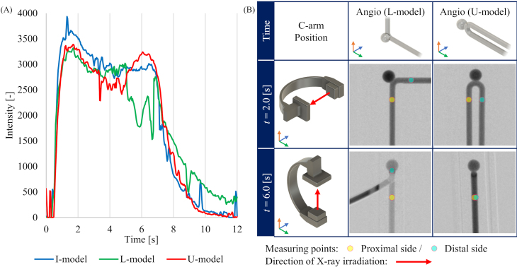
Objective: The aim of this study is to develop and validate a method for measuring the flow rate by data obtained from 3D-iFlow images - a prototype application in Four-dimensional digital subtraction angiography (4D-DSA).
Methods: Using phantom model and experimental circuit with circulating glycerin solution, an equation for the relationship between contrast media intensity and flow rate was developed. Applying the equation to the aneurysm phantom models, the derived flow rate was evaluated.
Results: The average errors between the derived flow rate and setting flow rate became larger when the glycerin flow and the X-rays from the X-ray tube of the angiography system were parallel to each other or when the measurement point included overlaps with other contrast enhanced areas.
Conclusion: Although the error increases dependent on the imaging direction and overlap of contrast enhanced area, the developed equation can estimate the flow rate using the image intensity value measured on 3D-iFlow based on 4D-DSA.
Keywords: Aneurysm; four-dimensional digital subtraction angiography; hemodynamics.
Conflict of interest statement
K. Otani is a full-time employee of Siemens Healthcare. The other authors have no conflicts of interest to report.
Figures


Similar articles
-
Dual-energy CT angiography in the evaluation of intracranial aneurysms: image quality, radiation dose, and comparison with 3D rotational digital subtraction angiography.AJR Am J Roentgenol. 2010 Jan;194(1):23-30. doi: 10.2214/AJR.08.2290. AJR Am J Roentgenol. 2010. PMID: 20028901
-
4D Digital Subtraction Angiography for the Temporal Flow Visualization of Intracranial Aneurysms and Vascular Malformations.J Stroke Cerebrovasc Dis. 2020 Dec;29(12):105327. doi: 10.1016/j.jstrokecerebrovasdis.2020.105327. Epub 2020 Sep 28. J Stroke Cerebrovasc Dis. 2020. PMID: 32992207
-
Quantification of Blood Velocity with 4D Digital Subtraction Angiography Using the Shifted Least-Squares Method.AJNR Am J Neuroradiol. 2018 Oct;39(10):1871-1877. doi: 10.3174/ajnr.A5793. Epub 2018 Sep 13. AJNR Am J Neuroradiol. 2018. PMID: 30213811 Free PMC article.
-
Quantitative and Qualitative Comparison of 4D-DSA with 3D-DSA Using Computational Fluid Dynamics Simulations in Cerebral Aneurysms.AJNR Am J Neuroradiol. 2019 Sep;40(9):1505-1510. doi: 10.3174/ajnr.A6172. AJNR Am J Neuroradiol. 2019. PMID: 31467234 Free PMC article.
-
4D-DSA: Development and Current Neurovascular Applications.AJNR Am J Neuroradiol. 2021 Jan;42(2):214-220. doi: 10.3174/ajnr.A6860. Epub 2020 Nov 26. AJNR Am J Neuroradiol. 2021. PMID: 33243899 Free PMC article. Review.
References
-
- Fujimura S, Yamanaka Y, Takao H, Ishibashi T, Otani K, Karagiozov K, et al. Hemodynamic and morphological differences in cerebral aneurysms between before and after rupture. J Neurosurg. 2023. Sep 1: 1-9. - PubMed
-
- Murayama Y, Fujimura S, Suzuki T, Takao H. Computational fluid dynamics as a risk assessment tool for aneurysm rupture. Neurosurg Focus. 2019. Jul 1; 47(1): E12. - PubMed
-
- Rajabzadeh-Oghaz H, van Ooij P, Veeturi SS, Tutino VM, Zwanenburg JJ, Meng H. Inter-patient variations in flow boundary conditions at middle cerebral artery from 7T PC-MRI and influence on Computational Fluid Dynamics of intracranial aneurysms. Comput Biol Med. 2020. May; 120: 103759. - PubMed
MeSH terms
Substances
LinkOut - more resources
Full Text Sources
Medical
