. 2024 Jul 23;43(7):114431.
doi: 10.1016/j.celrep.2024.114431. Epub 2024 Jul 4.

Elevating PLK1 overcomes BETi resistance in prostate cancer via triggering BRD4 phosphorylation-dependent degradation in mitosis

Affiliations

Elevating PLK1 overcomes BETi resistance in prostate cancer via triggering BRD4 phosphorylation-dependent degradation in mitosis

Yanquan Zhang et al. Cell Rep. .

Abstract

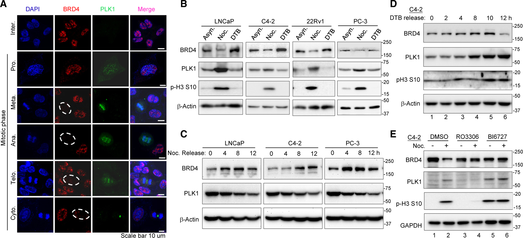
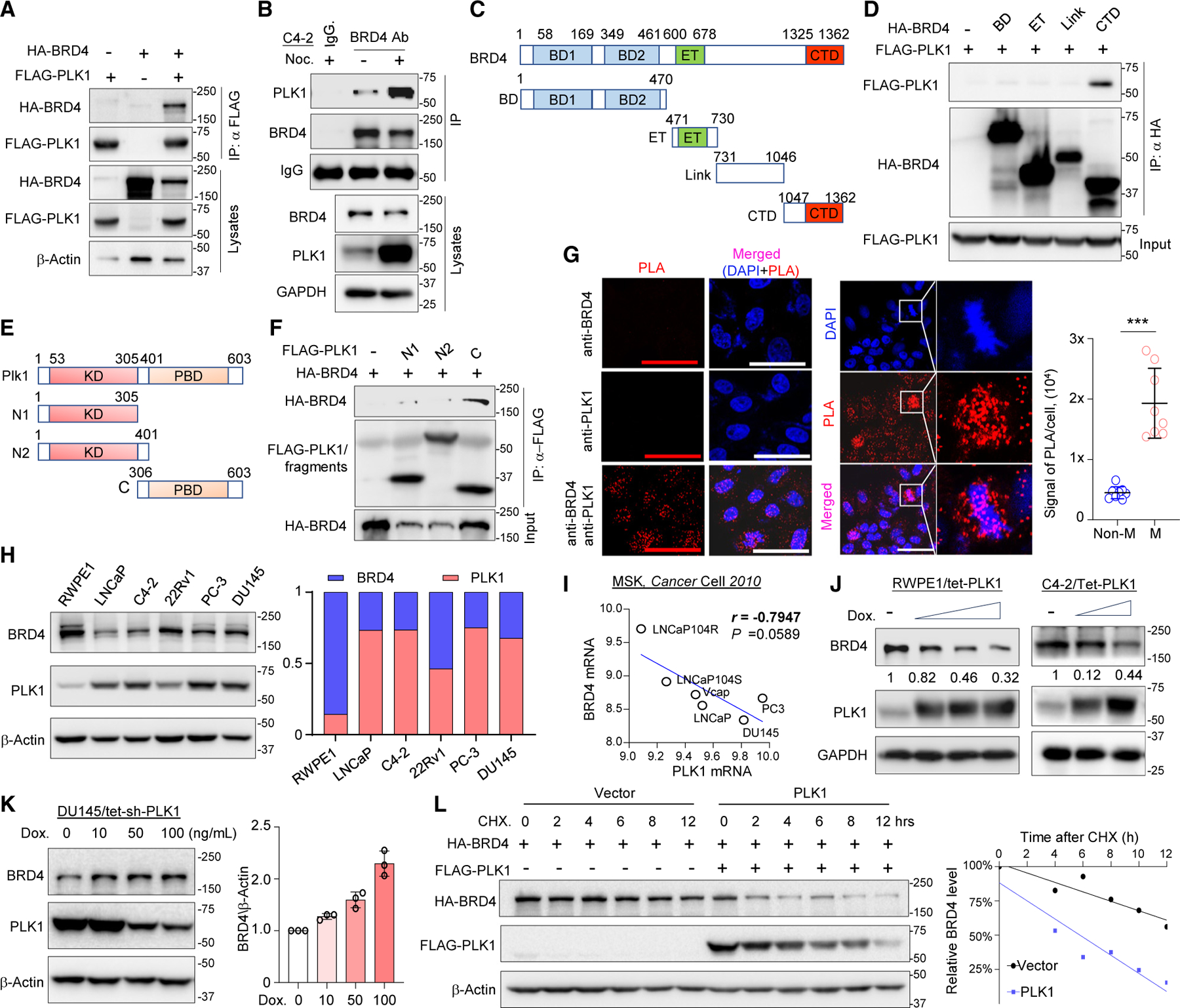
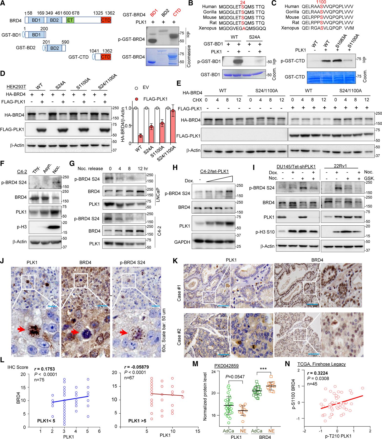
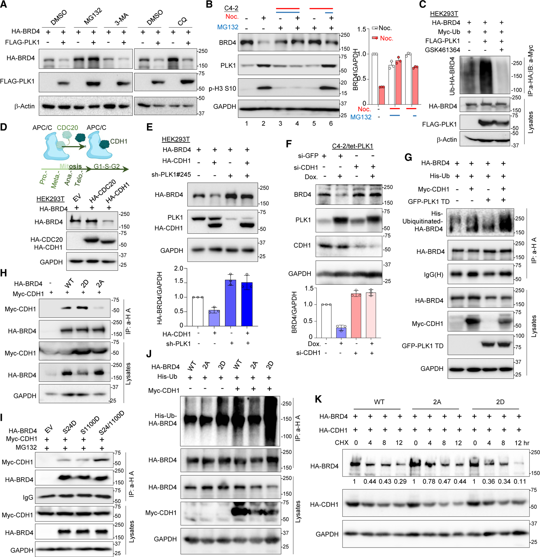
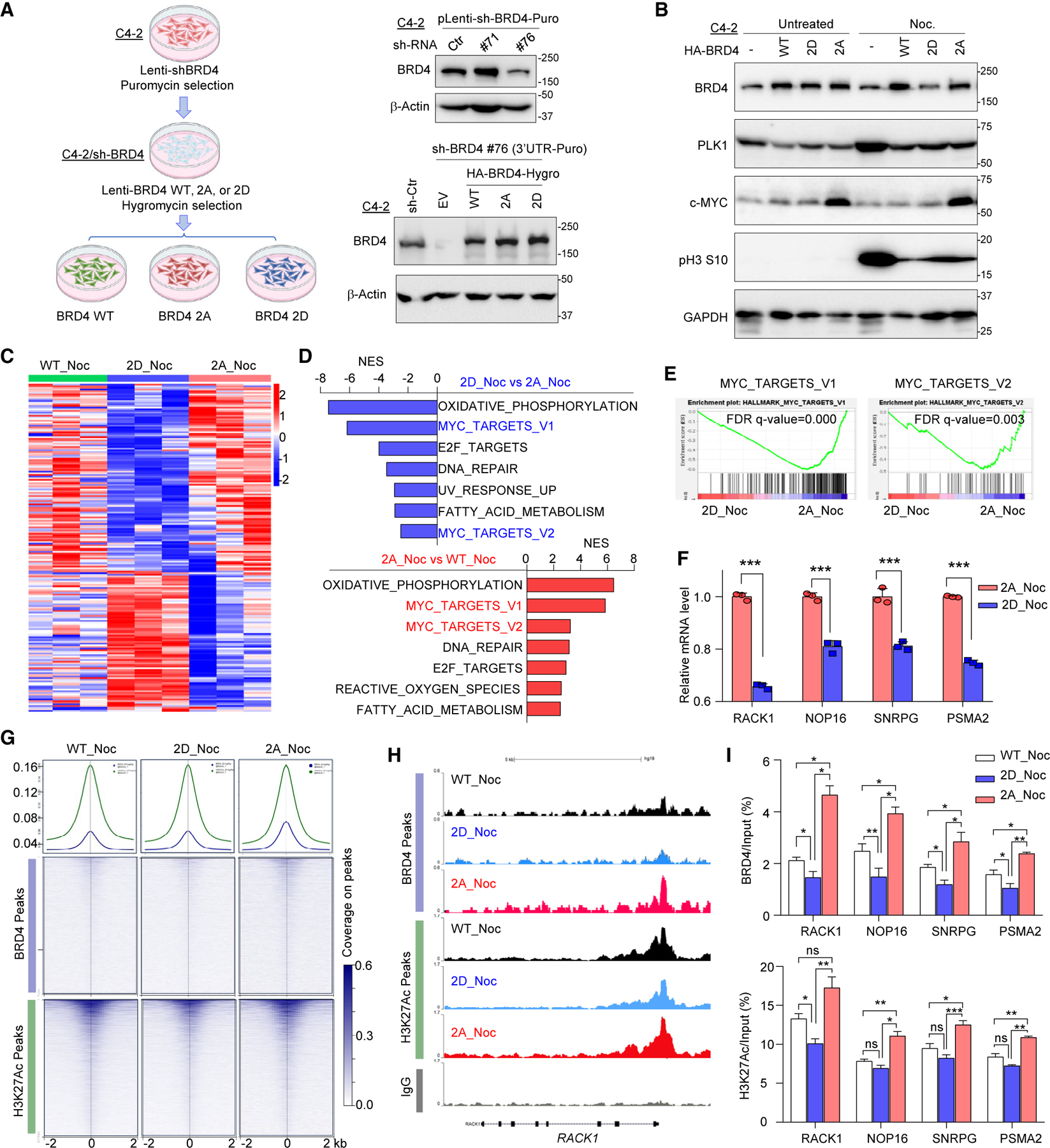
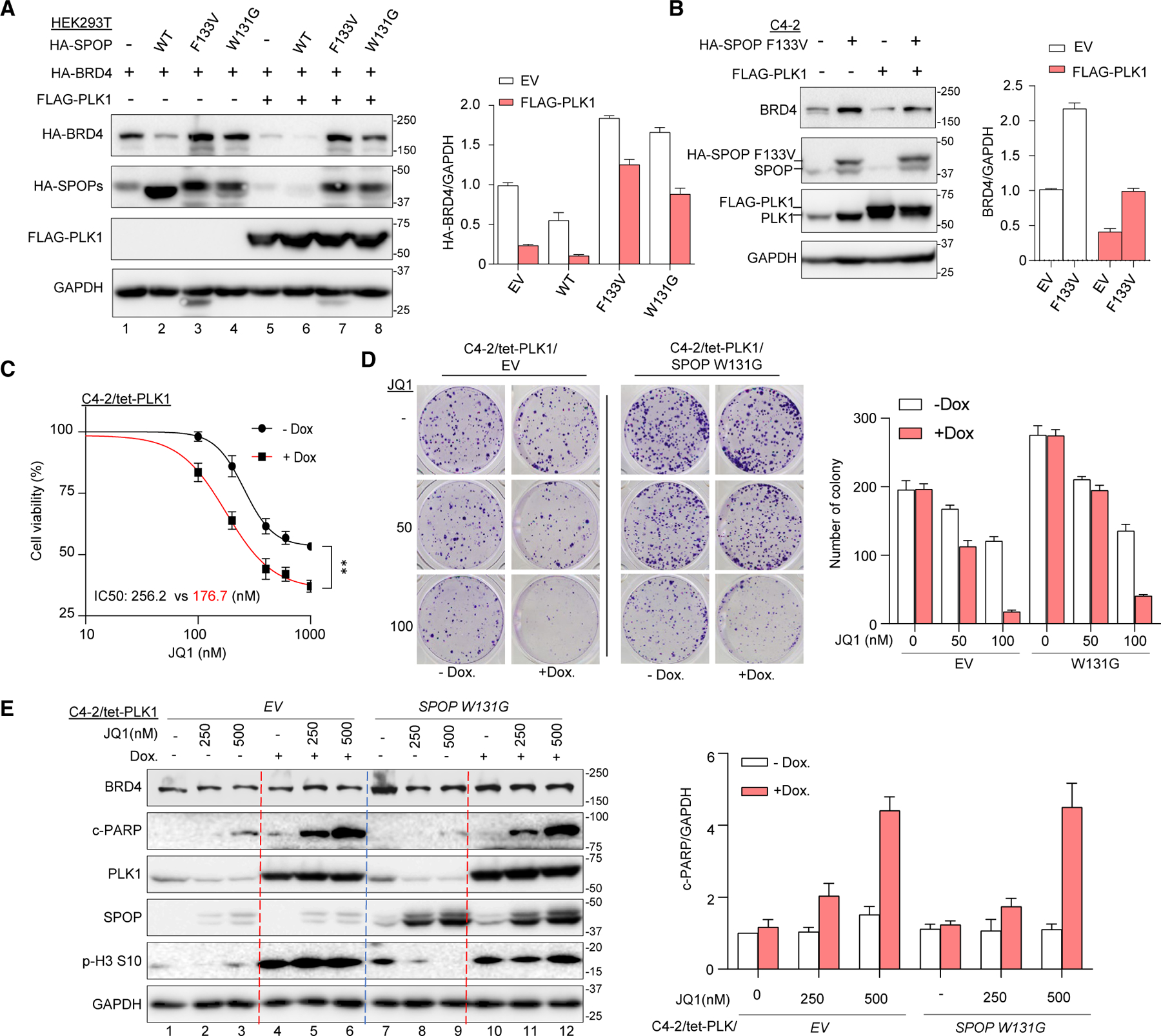
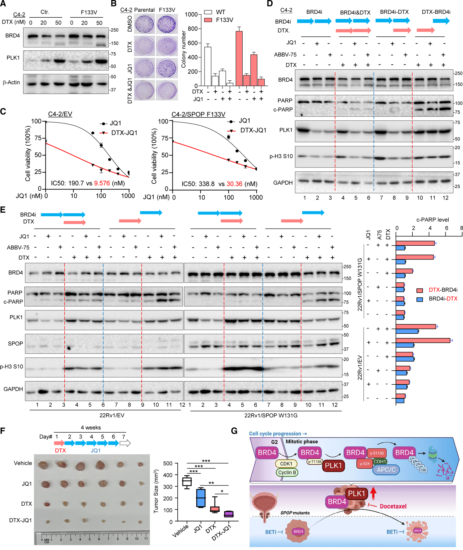
Bromodomain-containing protein 4 (BRD4) has emerged as a promising therapeutic target in prostate cancer (PCa). Understanding the mechanisms of BRD4 stability could enhance the clinical response to BRD4-targeted therapy. In this study, we report that BRD4 protein levels are significantly decreased during mitosis in a PLK1-dependent manner. Mechanistically, we show that BRD4 is primarily phosphorylated at T1186 by the CDK1/cyclin B complex, recruiting PLK1 to phosphorylate BRD4 at S24/S1100, which are recognized by the APC/CCdh1 complex for proteasome pathway degradation. We find that PLK1 overexpression lowers SPOP mutation-stabilized BRD4, consequently rendering PCa cells re-sensitized to BRD4 inhibitors. Intriguingly, we report that sequential treatment of docetaxel and JQ1 resulted in significant inhibition of PCa. Collectively, the results support that PLK1-phosphorylated BRD4 triggers its degradation at M phase. Sequential treatment of docetaxel and JQ1 overcomes BRD4 accumulation-associated bromodomain and extra-terminal inhibitor (BETi) resistance, which may shed light on the development of strategies to treat PCa.

Keywords: APC/C(Cdh1); BET inhibitor resistance; BRD4; CP: Cancer; CP: Molecular biology; PLK1; phosphorylation; prostate cancer.


Conflict of interest statement

Declaration of interests The authors declare no competing interests.

Figures

Figure 1.
Figure 1.. BRD4 is decreased during mitosis
(A) IF staining of C4-2 cells to detect BRD4 (red) and PLK1 (green). The white broken circles indicate mitotic cells. (B) WB analysis of PCa cells treated with DMSO, nocodazole (Noc.), or double-thymidine block (DTB). (C) WB of PCa cells released from nocodazole treatment. (D) WB of C4-2 cells released from DTB treatment. (E) WB of C4-2 cells upon indicated treatment.
Figure 2.
Figure 2.. PLK1 interacts with BRD4 and triggers its degradation
(A) Immunoprecipitation (IP) assay of HEK293T lysates with FLAG-PLK. (B) IP assay of C4-2 cell lysates with anti-BRD4 antibody. (C) The schematic structure of BRD4. (D) IP assay with HA-BRD4 fragments. (E) The schematic structure of PLK1. (F) IP assay using FLAG-PLK1 domains. (G) Proximity ligation assay (PLA) of BRD4 and PLK1 (left). Scale bar, 50 μm. The representative PLA images of metaphase cells are highlighted in the middle panel. The right histogram shows the quantified PLA signal as means ± SD of cells at M phase and at non-M phase. ***p < 0.001. (H) WB analysis of prostate non-transformed or cancer cell lines. The histogram on the right shows quantified BRD4 and PLK1. (I) The correlation analysis of BRD4 and PLK1 mRNA in PCa cell lines (GSE21032). (J) WB analysis of RWPE1 or C4-2/tet-inducible-PLK1 cells treated with doxycycline. (K) WB analysis of DU145/tet-shPLK1 cells treated with the indicated dosage of doxycycline. (L) WB of HEK293T cells transfected indicated plasmids upon cycloheximide treatment.
Figure 3.
Figure 3.. PLK1 phosphorylates BRD4 at S24 and S1100
(A) The left panel displays the schematic structure of GST-BRD4 fragments, while the right panel illustrates the result of an in vitro kinase assay. (B and C) In vitro kinase assays of purified GST-BRD4 WT or indicated alanine mutants. The upper panels display the conserved sequences of BRD4 among different species. (D) WB analysis of HEK293T cells transfected with plasmids indicated. The right panel indicates the quantified HA-BRD4 abundance. (E) WB of HEK293T cells transfected with plasmids indicated upon cycloheximide (CHX) treatment. (F) WB of C4-2 cells treated with thymidine (Thy.), nocodazole (Noc.), or DMSO (Asyn.). (G) WB of LNCaP (upper panel) and C4-2 (lower panel) cells released from nocodazole treatment. (H) WB of C4-2/tet-inducible PLK1 cells treated with increasing dosages of doxycycline. (I) WB of DU145/tet-shPLK1 cells upon doxycycline induction (left) or 22Rv1 cells upon PLK1 activity inhibition (right). (J) Representative images of IHC in human PCa samples. The red arrow indicates the same cell in serial slides. (K) Representative images of IHC against PLK1 or BRD4 in a TMA of PCa patients. (L) The correlation analysis between PLK1 and BRD4 based on the scores of (K). (M) The trend of PLK1 and BRD4 protein between adenocarcinoma and neuroendocrine PCa patients (ProteomeXchange #PXD042867). (N) The correlation analysis of pT210-PLK1 and pS1100-BRD4 using phosphoproteomic data of breast invasive carcinoma (TCGA, Firehose Legacy).
Figure 4.
Figure 4.. APC/CCdh1 complex promotes PLK1 phosphorylation-dependent BRD4 degradation
(A) WB of HEK293T cells transfected with HA-BRD4 and FLAG-PLK1 upon treatment of MG132, 3-MA, or chloroquine (CQ). (B) WB of C4-2 cells treated with nocodazole and MG132 in different manners. (C) Ubiquitination assay of HA-BRD4 with co-expression of FLAG-PLK1 and Myc-Ub. (D) The upper panel illustrates the APC/C complex working model during the cell cycle, while the lower panel shows WB analysis of HEK293T cells transfected with indicated plasmids. (E) WB of HEK293T cells transfected indicated plasmids, with the lower panel showing the relative HA-BRD4 amount. (F) WB of C4-2/tet-PLK1 cells transfected with indicated small interfering RNAs (siRNAs), with the lower panel showing the relative levels of endogenous BRD4. (G) Ubiquitination of HA-BRD4 with co-expression of indicated plasmids. (H) IP assay of HEK293T cells lysates transfected with indicated plasmids. (I) IP assay of Myc-CDH1 and HA-BRD4s. (J) Ubiquitination assay of HA-BRD4s with or without Myc-CDH1. (K) WB of HEK293T cells transfected with indicated plasmids upon CHX treatment, with the relative protein amounts of HA-BRD4s at each time point shown.
Figure 5.
Figure 5.. PLK1 phosphorylation of BRD4 inhibits its transcriptional activity
(A) Construction of C4-2 cells expressing different forms of BRD4s. (B) WB analysis of C4-2 expressing HA-BRD4s with/without nocodazole treatment. (C) Heatmap of top 200 DEGs (differentially expressed genes) among C4-2/BRD4s upon nocodazole treatment. (D) Top seven enriched GSEA-Hallmark pathways based on DEGs of RNA-seq. (E) Representative pathways MYC_tragets_V1 and V2 significantly enriched in GSEA Hallmark. (F) qPCR verification of Myc-targeted genes. (G) Intensity plots (upper) and heatmaps (lower) of coverage peaks of BRD4 and H3K27Ac at the C4-2/BRD4s cells genome with nocodazole treatment. (H) BRD4 and H3K27Ac binding peaks at the RACK1 promoter region of C4-2/BRD4s cells. (I) qPCR result of anti-BRD4 or anti-H3K27Ac chromatin immunoprecipitated DNA of C4-2/BRD4s cells. All data are shown as means ± SD from three times of independent assay. *p < 0.05, **p < 0.01, ***p < 0.001.
Figure 6.
Figure 6.. Ectopic expression of PLK1 overcomes SPOP-mutation-associated resistance to BRD4 inhibitors
(A) WB analysis of HEK293T cells transfected with indicated plasmids. The right panel shows the relative HA-BRD4 amount. (B) WB of C4-2/HA-SPOP-F133V and FLAG-PLK1 cells, with the right panel showing the relative endogenous BRD4. (C) The cell viability of C4-2/tet-PLK1 upon doxycycline and dosage of JQ1 treatment. **p < 0.01. (D) Representative colony images, with the quantity of each group on the right panel. (E) WB analysis of C4-2/tet-PLK1 and SPOP-W131G or control cells upon dosages of JQ1 and doxycycline treatment, with the right panel showing the relative levels of c-PARP.
Figure 7.
Figure 7.. Sequential treatment of docetaxel and BETi overcomes SPOP-mutation-associated resistance to BETi in PCa
(A) WB analysis of C4-2/SPOP-F133V or control cells treated with dosages of docetaxel (DTX). (B) The representative colony images of C4-2/SPOP-F133V or control cells treated with docetaxel and/or JQ1, with the quantity of each wells shown in the right panel. (C) The viability of C4-2/SPOP-F133V and control cells treated with docetaxel and dosages of JQ1. (D) WB analysis of C4-2 cells sequentially treated with BRD4 inhibitors and docetaxel in the indicated order. Each blue or red bar above represents 24 h. (E) WB analysis of 22Rv1/SPOP W131G or control cells sequentially treated with BRD4 inhibitors and docetaxel in the order shown, with the right panel showing the relative levels of c-PARP. (F) PCa patient-derived xenograft (PDX) tumors upon sequential treatment with docetaxel and JQ1, with the right panel showing the tumor sizes. *p < 0.05, **p < 0.01, ***p < 0.001. (G) A working model.

Similar articles

Cited by

References

    1. Wang CY, and Filippakopoulos P (2015). Beating the odds: BETs in disease. Trends Biochem. Sci 40, 468–479. 10.1016/j.tibs.2015.06.002. - DOI - PubMed
    1. Shi J, and Vakoc CR (2014). The Mechanisms behind the Therapeutic Activity of BET Bromodomain Inhibition. Mol. Cell 54, 728–736. 10.1016/j.molcel.2014.05.016. - DOI - PMC - PubMed
    1. Filippakopoulos P, and Knapp S (2014). Targeting bromodomains: epigenetic readers of lysine acetylation. Nat. Rev. Drug Discov 13, 337–356. 10.1038/nrd4286. - DOI - PubMed
    1. Duan Y, Guan Y, Qin W, Zhai X, Yu B, and Liu H (2018). Targeting Brd4 for cancer therapy: inhibitors and degraders. Medchemcomm 9, 1779–1802. 10.1039/c8md00198g. - DOI - PMC - PubMed
    1. Faivre EJ, McDaniel KF, Albert DH, Mantena SR, Plotnik JP, Wilcox D, Zhang L, Bui MH, Sheppard GS, Wang L, et al. (2020). Selective inhibition of the BD2 bromodomain of BET proteins in prostate cancer. Nature 578, 306–310. 10.1038/s41586-020-1930-8. - DOI - PubMed

Publication types

MeSH terms