. 2024 Sep 20;5(3):103164.
doi: 10.1016/j.xpro.2024.103164. Epub 2024 Jul 4.

Protocol for adeno-associated virus-mediated optogenetic activation of olfactory output neurons in neonatal mice

Affiliations

Protocol for adeno-associated virus-mediated optogenetic activation of olfactory output neurons in neonatal mice

Yu-Nan Chen et al. STAR Protoc. .

Abstract

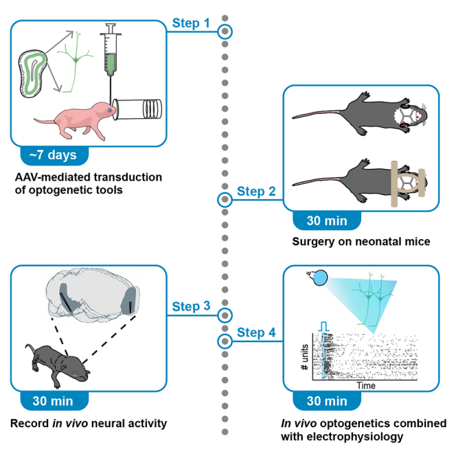
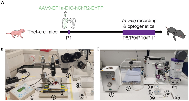
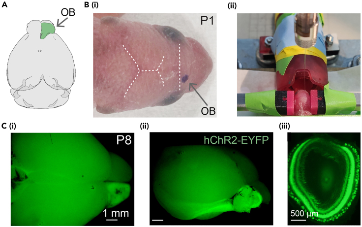
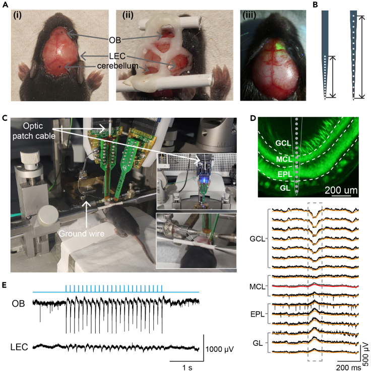
Optogenetic manipulation has proven a powerful tool for investigating the mechanisms underlying the function of neuronal networks, but implementing the technique on mammals during early development remains challenging. Here, we present a comprehensive workflow to specifically manipulate mitral/tufted cells (M/TCs), the output neurons in the olfactory circuit, mediated by adeno-associated virus (AAV) transduction and light stimulation in neonatal mice and monitor neuronal and network activity with in vivo electrophysiology. This method represents an efficient approach to elucidate functional brain development. For complete details on the use and execution of this protocol, please refer to Chen et al.1,2,3.

Keywords: biophysics; developmental biology; neuroscience; systems biology.


Conflict of interest statement

Declaration of interests The authors declare no competing interests.

Figures

None
Graphical abstract
Figure 1
Figure 1
Experimental design and setups (A) Experimental timeline. At P1, transduction of light-gated ChR2 channels in M/TCs was performed by AAV injections into the right OB of Tbet-cre mice. At P8, P9, P10 or P11, acute in vivo electrophysiological recordings were performed in the right OB combined with optogenetic activation of ChR2-expressing M/TCs. (B) The setup for microinjections on neonatal mice (devices corresponding to the numbers: ①infusion pump; ②stereotaxic apparatus; ③heating pad; ④anesthesia mask; ⑤stereomicroscope; ⑥ice box with viral construct; ⑦microliter syringe). (C) The setup for in vivo surgery (devices corresponding to the numbers: ⑧hot bead sterilizer; ⑨anesthesia induction box; ⑩anesthesia flow meter; ⑪isoflurane vaporizer; ⑫gas evacuation unit; ⑬isotonic sodium chloride solution; ⑭dental cement powder (left) and aqueous solution (right); ⑮needle; ⑯scissors; ⑰forceps).
Figure 2
Figure 2
Procedure for ChR2 transduction in the OB by virus injection at P1 (A) (i) 3D schematic of the OB position in the brain with the injected area in green (top view). (B) (i) An anesthetized P1 mouse with injection location for the right OB marked. (ii) A head-fixed P1 mouse in front of the anesthesia mask and the tip of the microliter syringe positioned into the right OB for viral injection. (C) Pictures showing hChR2-EYFP expression in the right OB (i: top view; ii: lateral view of the brain) from a P8 Tbet-cre mouse, and expression pattern across the OB layers on a coronal brain slice (iii).
Figure 3
Figure 3
Procedure for in vivo electrophysiological recording combined with optogenetics (A) Anesthetized neonatal mouse with recording locations marked (i) and exposed (ii) for multi-site extracellular recordings. The two plastic bars were mounted on the nasal and occipital bones respectively with the dental cement for tight head fixation during the recording. (iii) Overlaid photograph (from pictures taken under 440 nm blue light and neutral white light) showing the expression of hChR2-EYFP in the OB through the surface of the brain. (B) Schematic reconstruction of extracellular recording electrode tips with 50 μm (left) and 100 μm (right) inter-site spacing. Scale bars correspond to 750 μm (left) and 1500 μm (right) respectively. (C) Head-fixed neonatal mouse in the stereotaxic apparatus with optrode inserted in OB and electrode inserted in LEC. The optrode is attached to the laser delivery system (upper inner photo) for optogenetic stimulation. The silver wire is positioned into the cerebellum as ground and reference (lower inner photo). (D) Digital post-mortem photomontage reconstructing the location of the 16 recording channels of optrode in ventral OB of a P11 hChR2-EYFP expressing mouse (top; GCL: granule cell layer; MCL: mitral cell layer; EPL: external plexiform layer; GL: glomerular layer), and the laminar spontaneous activity across the corresponding OB layers (down; raw recording traces in black, 1–12 Hz band-pass filtered LFP traces in orange). Note that the 1–12 Hz LFPs reverse at the channel below the first MCL recording channel (traces in gray and red). (E) Representative traces of simultaneous extracellular recordings in OB and LEC with both LFPs and MUA during blue light (473 nm) pulse stimulation at 8 Hz. The TTL signals indicating 3-ms light square pulses are marked in light blue.
Figure 4
Figure 4
Effects of optogenetic stimulation on ChR2-expressing M/TCs with different light intensities (A) Averaged MCL field potentials induced by the first blue light (473 nm) pulse delivered to the OB of P8-11 mice with the light intensity set as 1%, 5% 10% and 20% of the maximal intensity (1.75 mW) of laser power. The corresponding light intensities measured at the tip of the optrode are indicated following the percentage. The 3-ms light square pulses are denoted by light blue bars. (B) Raster plots of OB neuronal firing in response to the first blue light pulse delivered in M/TCs of P8-11 mice at the corresponding intensity in (A).
Figure 5
Figure 5
LFP responses to light stimulation of ChR2-expressing M/TCs (A) (i) Averaged field potentials induced by 2 Hz blue light (473 nm) pulses in the MCL of OB from P8-11 mice. The 3-ms light square pulses are denoted by light blue bars. (ii) Color map of current source density (CSD) across 16 recording channels in the OB during the same periods as (i). (B) Averaged field potentials induced by blue light pulses delivered at 4 Hz (i)/8 Hz (ii)/16 Hz (iii)/32 Hz (iv) in the MCL of OB from P8-11 mice. Data are presented as median ± standard error of the median. (C) (i) Averaged peak amplitude of potentials induced by the first (black solid line) and second (black dashed line) light pulses at different inter-pulse intervals. (ii) Quantified paired-pulse ratio (2nd peak amplitude/1st peak amplitude). Data are presented as median ± standard error of the median. (D) Color maps of CSD in the OB in response to the first and second blue light pulses at 4 Hz, 8 Hz, 16 Hz and 32 Hz. The 3-ms light square pulses are denoted by light blue bars.
Figure 6
Figure 6
Neuronal firing responses to light stimulation of ChR2-expressing M/TCs (A) Averaged probability of neuronal firing in the OB and LEC of P8-11 mice in response to 3-ms blue light (473 nm) pulses delivered to the OB. The 3-ms light square pulses are denoted by light blue bars. Data are presented as median ± standard error of the median. (B) Z-scored firing rates of neurons in OB and LEC of P8-11 mice in response to 3-ms blue light pulses delivered to the OB.

Similar articles

References

    1. Chen Y.-N., Kostka J.K., Bitzenhofer S.H., Hanganu-Opatz I.L. Olfactory bulb activity shapes the development of entorhinal-hippocampal coupling and associated cognitive abilities. Curr. Biol. 2023;33:4353–4366.e5. doi: 10.1016/j.cub.2023.08.072. - DOI - PMC - PubMed
    1. Gretenkord S., Kostka J.K., Hartung H., Watznauer K., Fleck D., Minier-Toribio A., Spehr M., Hanganu-Opatz I.L. Coordinated electrical activity in the olfactory bulb gates the oscillatory entrainment of entorhinal networks in neonatal mice. PLoS Biol. 2019;17 doi: 10.1371/journal.pbio.2006994. - DOI - PMC - PubMed
    1. Kostka J.K., Hanganu-Opatz I.L. Olfactory-driven beta band entrainment of limbic circuitry during neonatal development. J. Physiol. 2023;601:3605–3630. doi: 10.1113/JP284401. - DOI - PubMed
    1. Schaal B., Saxton T.K., Loos H., Soussignan R., Durand K. Olfaction scaffolds the developing human from neonate to adolescent and beyond. Philos. Trans. R. Soc. Lond. B Biol. Sci. 2020;375 doi: 10.1098/rstb.2019.0261. - DOI - PMC - PubMed
    1. Miller S.S., Spear N.E. Olfactory learning in the rat neonate soon after birth. Dev. Psychobiol. 2008;50:554–565. doi: 10.1002/dev.20318. - DOI - PMC - PubMed

Publication types

LinkOut - more resources