

Characterizing T1 in the fetal brain and placenta over gestational age at 0.55T
- PMID: 38968093
- PMCID: PMC7617244
- DOI: 10.1002/mrm.30193
Characterizing T1 in the fetal brain and placenta over gestational age at 0.55T
Abstract
Purpose: T1 mapping and T1-weighted contrasts have a complimentary but currently under utilized role in fetal MRI. Emerging clinical low field scanners are ideally suited for fetal T1 mapping. The advantages are lower T1 values which results in higher efficiency and reduced field inhomogeneities resulting in a decreased requirement for specialist tools. In addition the increased bore size associated with low field scanners provides improved patient comfort and accessibility. This study aims to demonstrate the feasibility of fetal brain T1 mapping at 0.55T.
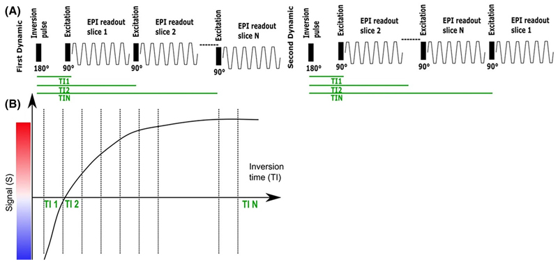
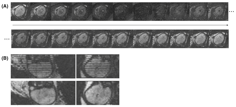
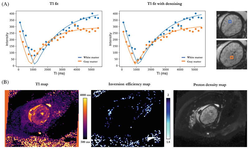
Methods: An efficient slice-shuffling inversion-recovery echo-planar imaging (EPI)-based T1-mapping and postprocessing was demonstrated for the fetal brain at 0.55T in a cohort of 38 fetal MRI scans. Robustness analysis was performed and placental measurements were taken for validation.
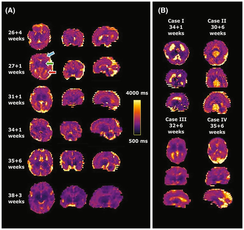
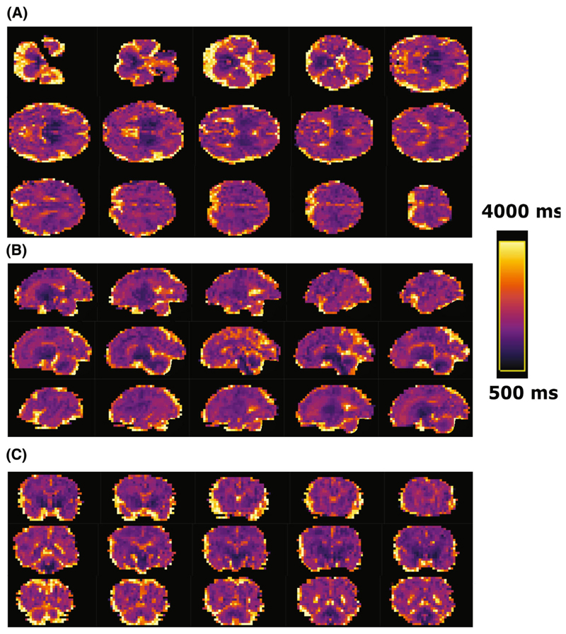
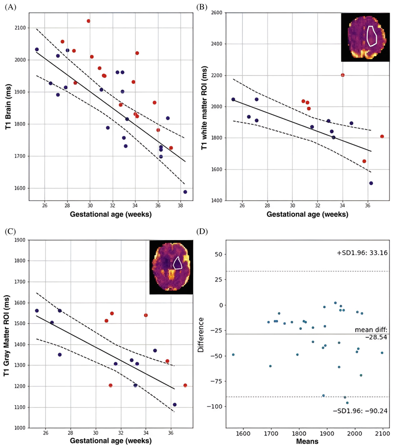
Results: High-quality T1 maps allowing the investigation of subregions in the brain were obtained and significant correlation with gestational age was demonstrated for fetal brain T1 maps ( ) as well as regions-of-interest in the deep gray matter and white matter.
Conclusions: Efficient, quantitative T1 mapping in the fetal brain was demonstrated on a clinical 0.55T MRI scanner, providing foundations for both future research and clinical applications including low-field specific T1-weighted acquisitions.
Keywords: fetal MRI; low cost; low‐field; relaxometry.
© 2024 The Author(s). Magnetic Resonance in Medicine published by Wiley Periodicals LLC on behalf of International Society for Magnetic Resonance in Medicine.
Figures

Similar articles
-
Real-time fetal brain and placental T2* mapping at 0.55T MRI.Magn Reson Med. 2025 Aug;94(2):615-624. doi: 10.1002/mrm.30497. Epub 2025 Mar 10. Magn Reson Med. 2025. PMID: 40065547 Free PMC article.
-
T2* relaxometry of fetal brain structures using low-field (0.55T) MRI.Magn Reson Med. 2025 May;93(5):1942-1953. doi: 10.1002/mrm.30409. Epub 2024 Dec 31. Magn Reson Med. 2025. PMID: 39737688 Free PMC article.
-
T1-weighted fast fluid-attenuated inversion-recovery sequence (T1-FFLAIR) enables the visualization and quantification of fetal brain myelination in utero.Eur Radiol. 2024 Jul;34(7):4573-4584. doi: 10.1007/s00330-023-10401-z. Epub 2023 Nov 29. Eur Radiol. 2024. PMID: 38019312 Free PMC article.
-
Magnetic resonance imaging of the fetal brain.Hong Kong Med J. 2016 Jun;22(3):270-8. doi: 10.12809/hkmj154678. Epub 2016 Apr 22. Hong Kong Med J. 2016. PMID: 27101791 Review.
-
Fetal and placental volumetric and functional analysis using echo-planar imaging.Top Magn Reson Imaging. 2001 Feb;12(1):52-66. doi: 10.1097/00002142-200102000-00006. Top Magn Reson Imaging. 2001. PMID: 11215716 Review.
Cited by
-
Real-time fetal brain and placental T2* mapping at 0.55T MRI.Magn Reson Med. 2025 Aug;94(2):615-624. doi: 10.1002/mrm.30497. Epub 2025 Mar 10. Magn Reson Med. 2025. PMID: 40065547 Free PMC article.
References
-
- Gowland PA, Freeman A, Issa B, et al. In vivo relaxation time measurements in the human placenta using echo planar imaging at 0.5 T. Magn Reson Imaging. 1998 Apr;16:241–247. - PubMed
-
- Aertsen M, Diogo MC, Dymarkowski S, Deprest J, Prayer D. Fetal MRI for dummies: what the fetal medicine specialist should know about acquisitions and sequences. Prenat Diagn. 2020;40:6–17. - PubMed
MeSH terms
Grants and funding
LinkOut - more resources
Full Text Sources
Medical
