. 2024 Nov 12;8(21):5557-5570.
doi: 10.1182/bloodadvances.2024012713.

Selective Btk inhibition by PRN1008/PRN473 blocks human CLEC-2, and PRN473 reduces venous thrombosis formation in mice

Affiliations

Selective Btk inhibition by PRN1008/PRN473 blocks human CLEC-2, and PRN473 reduces venous thrombosis formation in mice

Christopher W Smith et al. Blood Adv. .

Abstract

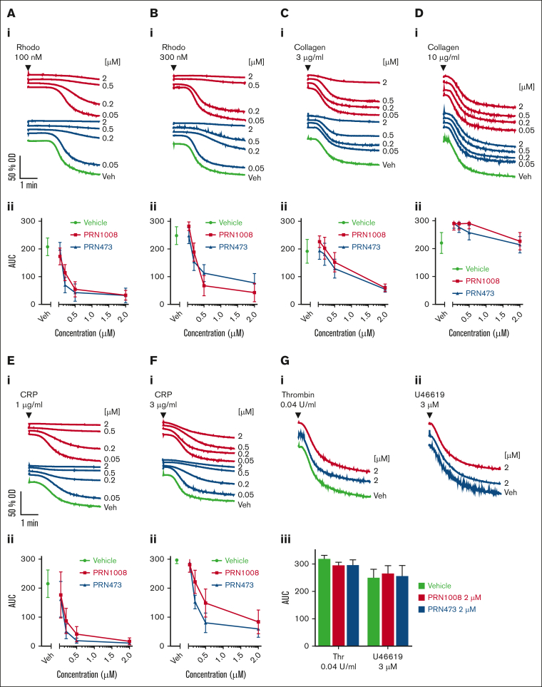
Platelet C-type lectin-like receptor 2 (CLEC-2) is a hem-immunoreceptor tyrosine-based activation motif-containing receptor that has a critical role in venous thrombosis but minimal involvement in hemostasis. CLEC-2 can be blocked by Btk inhibitors. Treatment with ibrutinib is associated with increased bleeding due to off-target inhibition of Src family kinases (SFKs). Patients with X-linked agammaglobulinemia (XLA) who lack Btk, however, do not bleed, suggesting selective Btk inhibition as a viable antithrombotic strategy. We assessed the effects of selective Btk inhibitors PRN1008 (rilzabrutinib) and PRN473 on platelet signaling and function mediated by CLEC-2 and glycoprotein-VI. We used healthy donors and XLA platelets to determine off-target inhibitor effects. Inferior vena cava (IVC) stenosis and Salmonella infection mouse models were used to assess antithrombotic effects of PRN473 in vivo. PRN1008 and PRN473 potently inhibited CLEC-2-mediated platelet activation to rhodocytin. No off-target inhibition of SFKs was seen. PRN1008 treatment of Btk-deficient platelets resulted in minor additional inhibition of aggregation and tyrosine phosphorylation, likely reflecting inhibition of Tec. No effect on G protein-coupled receptor-mediated platelet function was observed. PRN473 significantly reduced the number of thrombi in podoplanin-positive vessels after Salmonella infection and the presence of IVC thrombosis after vein stenosis. The potent inhibition of human platelet CLEC-2 and reduced thrombosis in in vivo models, together with the lack of off-target SFK inhibition and absence of bleeding reported in rilzabrutinib-treated patients with immune thrombocytopenia, suggest Btk inhibition as a promising antithrombotic strategy.


Conflict of interest statement

Conflict-of-interest disclosure: P.L.R.N., M.R.T., and S.P.W. have received research grants from Novartis, Principia Biopharma, and Rigel Pharmaceuticals. P.L.R.N. has received research funding from AstraZeneca and honoraria from Bayer, Grifols, and Takeda. The remaining authors declare no competing financial interests.

Figures

None
Graphical abstract
Figure 1.
Figure 1.
PRN1008 and PRN473 inhibit CLEC-2– and GPVI-mediated signaling. (A-B) Healthy donor–washed platelets (4 × 108/mL) were incubated with vehicle (0.02% DMSO) or indicated concentration (50, 200, 500, or 2000 nM) of Btk inhibitors PRN1008 and PRN473 for 1 hour, then stimulated with snake venom toxin rhodocytin 300 nM (A) or CRP 10 μg/mL (B) for 180 seconds in the presence of eptifibatide (9 μM) and lysed with reducing sample buffer. Whole cell lysates were then separated by sodium dodecyl sulfate–polyacrylamide gel electrophoresis (SDS-PAGE) and western blotted for tyrosine phosphorylation of indicated proteins. Total LAT was used as a loading control. Representative western blots (i) and normalized densitometry quantification (ii). Mean ± standard error of the mean (SEM) of 4 identical experiments. Bas, basal; Veh, vehicle; pan-pY, panphosphotyrosine.
Figure 2.
Figure 2.
PRN1008 and PRN473 inhibit CLEC-2– and GPVI–mediated platelet aggregation. Healthy donor–washed platelets (2 × 108/mL) were incubated with vehicle (0.02% DMSO) or indicated concentration (50, 200, 500, or 2000 nM) of Btk inhibitors PRN1008 and PRN473 for 1 hour before platelet aggregation to snake venom toxin rhodocytin 100 (A) and 300 nM collagen (B); 3 (C) and 10 μg/mL CRP (D); 1 (E) and 3 μg/mL (F) or thrombin (0.04 U/mL) and thromboxane A2 mimetic U46619 (3 μM) (G) were measured by lumi-aggregometry. Representative traces (i) and quantification (ii). Mean ± SEM (n = 6). OD, optical density; Rhodo, rhodocytin; Veh, vehicle.
Figure 3.
Figure 3.
PRN1008 inhibits CLEC-2– and GPVI–mediated platelet activation and CLEC-2–induced thrombus formation in whole blood. Citrated healthy donor whole blood was incubated with vehicle (0.02% DMSO) or indicated concentration (0.2, 0.5, 2, or 5 μM) of Btk inhibitors PRN1008 and PRN473 for 1 hour. Platelet activation, indicated by activated integrin αIIbβ3 (PAC-1) and P-selectin surface expression, was then assessed by flow cytometry in response to stimulation with snake venom toxin rhodocytin (100 and 300 nM) (A) or CRP (3 and 10 μg/mL) (B) or thrombin receptor activating peptide (TRAP; 30 μM and 100 uM) (C). Mean ± SEM (n = 4-8). (D) Citrated healthy donor whole blood was incubated with vehicle (0.02% DMSO) or indicated concentration (0.5 or 5 μM) of Btk inhibitors PRN1008 or PRN473 for 1 hour. Blood was then labeled with DiOC6 dye (10 minutes) and perfused over recombinant podoplanin-coated (10 μg/mL) channels at 150 per second for 8 minutes. Representative images (i); quantification of platelet surface area coverage (ii, iv); and mean aggregate size in images captured every 10 seconds (iii, v) are shown. Mean ± SEM (n = 3-4 per condition). Scale bar, 100 μm. Statistical analysis by 2-way analysis of variance (ANOVA) with Tukey correction for multiple comparisons. ∗P < .05; comparison with vehicle indicated by color.
Figure 4.
Figure 4.
PRN1008 blocks GPVI-mediated aggregation and PLCɣ2 phosphorylation in XLA platelets at low agonist concentrations of agonist. (A-D) Healthy donor and XLA washed platelets (2 × 108/mL) were incubated with vehicle (0.02% DMSO) or indicated concentration (0.5 and 5 μM) of PRN1008 for 1 hour before platelet aggregation to snake venom toxin rhodocytin 300 nM (A), collagen 3 (B) and 10 μg/mL (C), and CRP (3 μg/mL) (D) was measured by lumi-aggregometry. Representative traces (i) and quantification (ii); n = 3 for healthy donor; n = 2 for XLA. (E-F) Healthy donor and XLA washed platelets (4 × 108/mL) were incubated with vehicle (0.02% DMSO) or indicated concentration (0.5 or 5 μM) of PRN1008 for 1 hour, then stimulated with snake venom toxin rhodocytin 300 nM (E) or CRP 10 μg/mL (F) for 180 seconds in the presence of eptifibatide (9 μM) and lysed with reducing sample buffer. Whole cell lysates were then separated by SDS-PAGE and western blotted for tyrosine phosphorylation of indicated proteins. Total LAT was used as a loading control. Representative western blots (n = 1). OD, optical density; Veh, vehicle.
Figure 5.
Figure 5.
PRN1008 and PRN473 block platelet activation in mouse whole blood. (A) Mouse washed platelets (2 × 108/mL) were incubated with vehicle (0.02% DMSO) or indicated concentration (0.5, 2, 5, or 10 μM) of Btk inhibitors PRN1008 for 1 hour. Platelet activation, indicated by activated integrin αIIbβ3 (JON/A) and P-selectin surface expression, was then assessed by flow cytometry in response to stimulation with snake venom toxin rhodocytin 100 (Ai) and 300 nM (Aii) or CRP 3 (Bi) and 10 μg/mL (Bii) or PAR4 receptor activating peptide (100 or 500 nM) (C). Mean ± SEM (n = 3-6). Rhodo, rhodocytin.
Figure 6.
Figure 6.
PRN473 reduces Salmonella-induced liver thrombosis in mice. (A-B) WT mice were fed control or PRN473 formulated diet for 7 days, then had spleens harvested for Btk occupancy analysis (A) or citrated whole blood taken for platelet activation analysis by flow cytometry (B), indicated by activated integrin αIIbβ3 (JON/A) and P-selectin surface expression, after stimulation with snake venom toxin rhodocytin (100 and 300 nM), CRP (3 and 10 μg/mL), or PAR4 receptor activating peptide (500 nM). Mean ± SEM (n = 3). Statistical analysis by 2-way ANOVA with Sídák correction for multiple comparisons. (C) WT mice fed control or PRN473 formulated diet were infected with 5 × 105 CFU S typhimurium on day 7, with thrombi in portal vein assessed at day 14 (7 days after infection). Representative immunohistochemistry staining of frozen liver sections (scale bar, 200 μm) (i) and quantification of thrombus area per unit vessel area (ii), quantification of number of thrombi (iii), quantification of number of podoplanin-expressing vessels (iv), number of thrombi in podoplanin-positive levels (v) (n = 6). (D) Peripheral blood counts of S typhimurium–infected mice at baseline and 7 days after infection. Mean ± SEM (n = 4). (A-B) ∗P < .05; ∗∗P < .01; ∗∗∗∗P < .0001. CFU, colony forming units; WT, wild type.
Figure 7.
Figure 7.
PRN473 inhibits IVC thrombosis in mice. (A) WT mice were gavaged daily with vehicle or PRN473 (80 mg/kg) for 4 days, then had platelet activation measured in citrated whole blood by flow cytometry, as indicated by activated integrin αIIbβ3 (JON/A) and P-selectin surface expression, after stimulation with snake venom toxin rhodocytin (100 and 300 nM), CRP (3 and 10 μg/mL), or thrombin (0.03 and 0.1 U/mL). GPRP was added to thrombin stimulation to prevent fibrin clot formation. Mean ± SEM (n = 3). Statistical analysis by 2-way ANOVA with Sídák correction for multiple comparisons. (B) IVC stenosis model of DVT was performed on WT mice gavaged with vehicle or PRN473 (80 mg/kg) for 4 days. Mice were euthanized 6 hours after IVC ligation and had spleens harvested for Btk occupancy analysis (i) and citrated plasma taken for drug concentration measurements (ii), and assessment for thrombus prevalence (iii), length (iv), and weight (v). Median: n = 16-18 mice per condition for splenic Btk occupancy and thrombus analysis; n = 17-21 mice per condition for plasma drug concentration measurements. Statistical analysis by Mann-Whitney test. ∗P < .01; ∗∗∗P = .0002; ∗∗∗∗P < .0001. GPRP, Gly-Pro-Arg-Pro; Veh, vehicle; WT, wild type.

References

    1. Campos J, Ponomaryov T, De Prendergast A, et al. Neutrophil extracellular traps and inflammasomes cooperatively promote venous thrombosis in mice. Blood Adv. 2021;5(9):2319–2324. - PMC - PubMed
    1. Villa E, Cammà C, Marietta M, et al. Enoxaparin prevents portal vein thrombosis and liver decompensation in patients with advanced cirrhosis. Gastroenterology. 2012;143(5):1253–1260.e4. - PubMed
    1. Hitchcock JR, Cook CN, Bobat S, et al. Inflammation drives thrombosis after Salmonella infection via CLEC-2 on platelets. J Clin Invest. 2015;125(12):4429–4446. - PMC - PubMed
    1. Payne H, Ponomaryov T, Watson SP, Brill A. Mice with a deficiency in CLEC-2 are protected against deep vein thrombosis. Blood. 2017;129(14):2013–2020. - PMC - PubMed
    1. Chauhan A, Sheriff L, Hussain MT, et al. The platelet receptor CLEC-2 blocks neutrophil mediated hepatic recovery in acetaminophen induced acute liver failure. Nat Commun. 2020;11(1):1939. - PMC - PubMed

Publication types

MeSH terms

Supplementary concepts