. 2024 Nov:515:67-78.
doi: 10.1016/j.ydbio.2024.06.021. Epub 2024 Jul 3.

Planarian LDB and SSDP proteins scaffold transcriptional complexes for regeneration and patterning

Affiliations

Planarian LDB and SSDP proteins scaffold transcriptional complexes for regeneration and patterning

Taylor Medlock-Lanier et al. Dev Biol. 2024 Nov.

Abstract

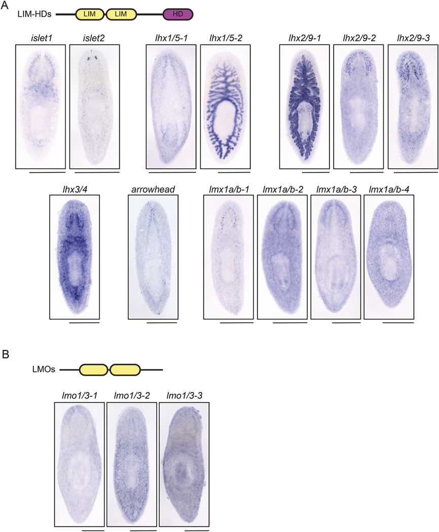
Sequence-specific transcription factors often function as components of large regulatory complexes. LIM-domain binding protein (LDB) and single-stranded DNA-binding protein (SSDP) function as core scaffolds of transcriptional complexes in animals and plants. Little is known about potential partners and functions for LDB/SSDP complexes in the context of tissue regeneration. In this work, we find that planarian LDB1 and SSDP2 promote tissue regeneration, with a particular function in anterior regeneration and mediolateral polarity reestablishment. We find that LDB1 and SSDP2 interact with one another and with characterized planarian LIM-HD proteins Arrowhead, Islet1, and Lhx1/5-1. We also show that SSDP2 and LDB1 function with islet1 in polarity reestablishment and with lhx1/5-1 in serotonergic neuron maturation. Finally, we find new roles for LDB1 and SSDP2 in regulating gene expression in the planarian intestine and parenchyma; these functions are likely LIM-HD-independent. Together, our work provides insight into LDB/SSDP complexes in a highly regenerative organism. Further, our work provides a strong starting point for identifying and characterizing potential binding partners of LDB1 and SSDP2 and for exploring roles for these proteins in diverse aspects of planarian physiology.

Keywords: Flatworm; Gene Expression; LDB; Planarian; Regeneration; SSDP; Schmidtea; Transcription factor.


Figures

Fig. 1.
Fig. 1.. Planarian SSDP and LDB homologs promote regeneration.
A) Diagram showing domain architecture in canonical LDB transcriptional complexes (Bronstein et al., 2010; Enkhmandakh et al., 2006). LDB = LIM Domain Binding protein; SSDP=Single-Stranded DNA Binding Protein; and LIM-HD = LIM-Homeodomain, where LIM is a region of homology first identified between Lin-11, Isl-1 and Mec-3. LDB proteins have a long dimerization domain (blue) and a LIM-binding domain (green). SSDP proteins have a conserved domain (red) and a long, unstructured region. LIM-HD proteins have a pair of LIM domains (yellow) and a homeodomain (purple). Complexes form to bind DNA and regulate gene expression. B) in situ hybridization showing expression patterns of planarian LDB1, LDB2, SSDP1, and SSDP2. All four genes are broadly expressed with some enrichment, particularly for LDB1 and SSDP2, in the brain. C) Images of live planarians after RNAi, head amputation, and 6 days of regeneration. control(RNAi) animals have a rounded blastema with two eyespots visible (n = 10). LDB1(RNAi) and SSDP2 (RNAi) regenerates have pointed blastemas with zero or one eyespot (6/6 and 9/10, respectively). D). in situ hybridization to mark the broadly expressed transcript ChAT (choline acetyltransferase (Nishimura et al., 2010);) in animals after RNAi, head amputation, and 6 days of regeneration. LDB1(RNAi) and SSDP2(RNAi) animals have smaller brains which are fused at the midline. E) Quantification of animals from a repetition of the experiment in (D) showing significantly smaller brains after LDB1(RNAi) and SSDP2(RNAi). (n = 5–9; Kruskal-Wallis with Dunn’s correction for multiple comparisons.) P is an adjusted value, * indicates P ≤ 0.05 and ** indicates P ≤ 0.005. Mean and SD are shown. Scale = 500 μm.
Fig. 2.
Fig. 2.. Planarian LDB1 interacts with SSDP2, as well as the LIM domains of Arrowhead, Islet, and Lhx1/5–1.
A) Diagram showing domain architecture for LDB1, SSDP2, Arrowhead, Islet, and Lhx1/5–1. “FL” indicates full length protein and “Sh” indicates a shorter truncation used in subsequent analyses. The color scheme used is the same as in Fig. 1A. B) Yeast two-hybrid results showing the interaction of full-length LDB1 with full-length SSDP2, Arrowhead, Islet, and Lhx1/5–1. Full-length Ssdp2, Islet, and Lhx1/5–1 exhibited transactivation when used as the Bait (dark grey), so these pairings were not used for the final assessment. Full length planarian LDB1 did not show interaction with itself, despite known homodimerization in other models. (- indicates no interaction; ± indicates weak growth; +, ++, or +++ indicates growth on restrictive medium). C) Yeast two-hybrid results showing that the core domains of LDB1 (LDB1-Sh) interact with the conserved domain of SSDP1 and the LIM domains of Arrowhead, Islet1, and Lhx1/5–1. In this experiment, we did note a weak self-interaction between the core domains of LDB1, which could support some dimerization of the protein. S. pombe Mob1 and Sid2 were used as controls (Balasubramanian et al., 1998; Hou et al., 2004). Cell growth for B and C is shown in Supp. Figs. 1A–B. D) Double FISH showing that LDB1 is coexpressed with SSDP2, arrowhead, islet1, and lhx1/51. Scale = 10 μm.
Fig. 3.
Fig. 3.. SSDP2(RNAi) and LDB1(RNAi) phenotypes indicate functional overlap with Islet1 and Lhx1/5–1.
A) Diagram depicting published expression patterns of planarian LIM-HD-encoding genes islet1 (Hayashi et al., 2011; Marz et al., 2013), arrowhead (Roberts-Galbraith et al., 2016), and lhx1/51 (Currie and Pearson, 2013). B) control(RNAi) and lhx1/51(RNAi) animals underwent in situ hybridization with TPH (tryptophan hydroxylase (Nishimura et al., 2007);) 6 days after head amputation. Fewer peripheral TPH+ neurons are seen after lhx1/51(RNAi) (see inset at bottom), as seen in a prior study (Currie and Pearson, 2013). C) control (RNAi), SSDP2(RNAi), LDB1(RNAi), and islet1(RNAi) animals stained for TPH+ via in situ hybridization 6 days after head amputation. Fused eyespots are visible after SSDP2(RNAi), LDB1(RNAi), or islet1(RNAi). Decreased peripheral TPH+ puncta were noted after knockdown of SSDP2 or LDB1, but not islet1 (see insets). D) control (RNAi), islet1(RNAi), SSDP2(RNAi) and LDB1(RNAi) animals underwent tail amputation, 6 days of regeneration, and in situ hybridization with the Wnt1 probe (Adell et al., 2009; Petersen and Reddien, 2008). Only posterior ends are shown. E) control(RNAi), islet1(RNAi), SSDP2(RNAi) and LDB1(RNAi) animals underwent head amputation, 7 days of regeneration, and in situ hybridization with the sFRP-1 probe (Gurley et al., 2008; Petersen and Reddien, 2008). Only anterior ends are shown. F) control(RNAi), islet1(RNAi), SSDP2(RNAi) and LDB1(RNAi) animals underwent head amputation, 6 days of regeneration, and in situ hybridization with the slit1 probe (Cebrià et al., 2007). Animals were imaged with the dorsal side facing up; arrowheads indicate the anterior-most dorsal slit signal. G) Diagrams of the amputation plane used for experiments in (H). H) control(RNAi), islet1(RNAi), SSDP2(RNAi) and LDB1(RNAi) animals underwent sagittal amputation, 9 days of regeneration, and in situ hybridization with the ChAT probe (Nishimura et al., 2010). Animals are shown with the regenerating side to the right. I) We measured the area of both the newly regenerated brain lobe (right) and the existing brain lobe (left) and created a ratio for each animal in (H). SSDP2(RNAi) and LDB1(RNAi) animals regenerated significantly less new brain tissue than control(RNAi) animals. (n = 6–11; Kruskal-Wallis with Dunn’s correction for multiple comparisons. P is an adjusted value, ** indicates P ≤ 0.005 and **** indicates P ≤ 0.0001). Mean and SD are shown. Scale = 500 μm (B, C, F, H), 100 μm (B, C insets), 200 μm (D, E).
Fig. 4.
Fig. 4.. Expression of planarian LIM-homeodomain and LIM-only-encoding genes.
A) Domain architecture of a typical LIM-homeodomain (LIM-HD) protein. 13 planarian genes encode LIM-HD proteins. Expression of these genes is shown in untreated planarians and genes are grouped by family. B) LIM-only (LMO) proteins have a typical domain architecture with two LIM domains each. 3 planarian genes encode LMO proteins. in situ hybridization for each LMO gene is shown. Scale = 500 μm.
Fig. 5.
Fig. 5.. Identification of genes differentially expressed after SSDP2(RNAi).
A) RNAi paradigm used for bulk RNA-seq experiments. Animals underwent head amputation and 6 days of regeneration. B) We identified 64 genes that were upregulated ≥1.5x with a P value < 0.05 after false discovery rate correction. We also identified 53 genes that were downregulated ≥1.5x with a P value < 0.05 after false discovery rate correction. C) In situ hybridization showing normal expression of four genes significantly downregulated after SSDP2(RNAi). Two parenchymal- and two intestine-enriched genes are shown. D) One transcript downregulated after SSDP2(RNAi) in our RNA-Seq data is dd_215. Using in situ hybridization, we show that dd_215 is absent from the blastema and is downregulated in both the existing intestine and pharynx after either LDB1(RNAi) or SSDP2(RNAi), but not after knockdown of the gut-enriched LIM-HD-encoding genes lhx1/52 or lhx2/91. E-H) dd_120 and dd_240 are decreased in expression after either LDB1(RNAi) or SSDP2(RNAi), but not after knockdown of the parenchymal-enriched lmx1a/b-4. Ventral (E–F) and dorsal (G–H) views of animals are shown. For dd_120, we noted expression in two distinct cell types with strong (arrowhead) and weak (arrow) expression. Cells with weak expression are nearly absent after LDB1(RNAi) or SSDP2(RNAi). Animals are 6 days post-head amputation and both ventral (top) and dorsal (bottom) views are shown. Scale = 200 μm.

Update of

References

    1. Adell T, Saló E, Boutros M, Bartscherer K, 2009. Smed-Evi/Wntless is required for beta-catenin-dependent and -independent processes during planarian regeneration. Development 136, 905–910. - PubMed
    1. Adler CE, Seidel CW, McKinney SA, Sánchez Alvarado A, 2014. Selective amputation of the pharynx identifies a FoxA-dependent regeneration program in planaria. Elife 3, e02238. - PMC - PubMed
    1. Agulnick AD, Taira M, Breen JJ, Tanaka T, Dawid IB, Westphal H, 1996. Interactions of the LIM-domain-binding factor Ldb1 with LIM homeodomain proteins. Nature 384, 270–272. - PubMed
    1. Balasubramanian MK, McCollum D, Chang L, Wong KC, Naqvi NI, He X, Sazer S, Gould KL, 1998. Isolation and characterization of new fission yeast cytokinesis mutants. Genetics 149, 1265–1275. - PMC - PubMed
    1. Becker T, Ostendorff HP, Bossenz M, Schluter A, Becker CG, Peirano RI, Bach I, 2002. Multiple functions of LIM domain-binding CLIM/NLI/Ldb cofactors during zebrafish development. Mech. Dev. 117, 75–85. - PubMed

Publication types

MeSH terms

LinkOut - more resources