

Spatially resolved imaging of human macular capillaries using adaptive optics-enhanced optical coherence tomography angiography
- PMID: 38969668
- PMCID: PMC11226425
- DOI: 10.1038/s41598-024-65534-y
Spatially resolved imaging of human macular capillaries using adaptive optics-enhanced optical coherence tomography angiography
Abstract
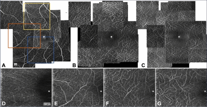
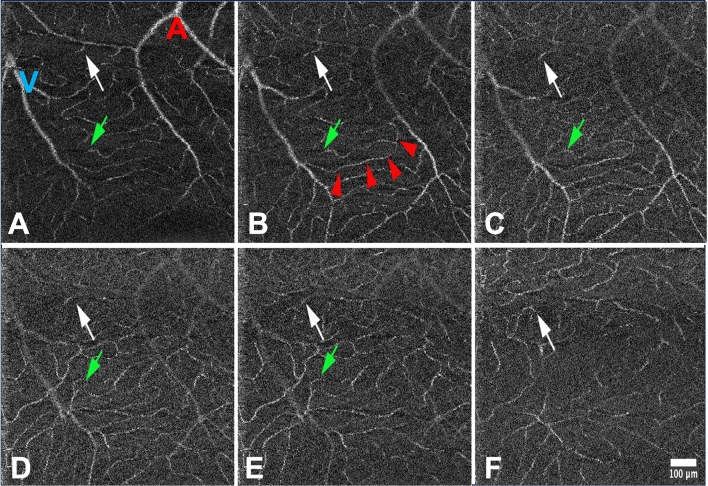
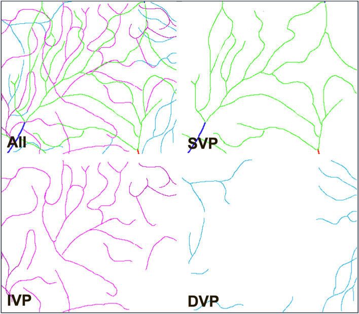
Documenting the organization of the retinal capillaries is of importance to understand the visual consequences of vascular diseases which may differentially affect the microvascular layers. Here we detailed the spatial organization of the macular capillaries in ten healthy human subjects using a prototypic adaptive optics-enhanced optical coherence tomography angiography (AO-OCTA) system. Within the central 6° × 6°, the radial peripapillary capillaries and the superficial, intermediate and deep vascular plexuses (SVP, IVP and DVP, respectively) were consistently resolved. In 8 out of the 10 eyes, the capillary segments composing the perifoveal arcade (PFA) were perfused only by the SVP, while drainage of the PFA showed more variability, comprising a case in which the PFA was drained by the DVP. Around the center, a distinct central avascular zone could be documented for each layer in 7 of the 10 cases; in three eyes, the IVP and SVP merged tangentially around the center. In all eyes, the foveal avascular zone was larger in the DVP than in the SVP and IVP. In one eye with incomplete separation of the inner foveal layers, there was continuity of both the SVP and the IVP; a central avascular zone was only present in the DVP. The diversity of perfusion and drainage patterns supported a connectivity scheme combining parallel and serial organizations, the latter being the most commonly observed in perifoveal vessels. Our results thus help to further characterize the diversity of organization patterns of the macular capillaries and to robustly analyze the IVP, which will help to characterize early stages of microvascular diseases.
Keywords: Fovea; Macula; Microcirculation; Optical coherence tomography angiography; Retina.
© 2024. The Author(s).
Conflict of interest statement
The authors declare that the research was conducted in the absence of any commercial or financial relationships that could be considered as a potential conflict of interest for this work. The corresponding author is responsible for submitting a competing interest statement on behalf of all authors.
Figures


References
-
- Balaratnasingam C, An D, Hein M, Yu P, Yu D-Y. Studies of the retinal microcirculation using human donor eyes and high-resolution clinical imaging: Insights gained to guide future research in diabetic retinopathy. Prog. Retin. Eye Res. 2023;94:101134. doi: 10.1016/j.preteyeres.2022.101134. - DOI - PubMed
MeSH terms
LinkOut - more resources
Full Text Sources
