Review
. 2024 Jul 5;15(1):5661.
doi: 10.1038/s41467-024-49949-9.

Neuroimaging and fluid biomarkers in Parkinson's disease in an era of targeted interventions

Affiliations
Review

Neuroimaging and fluid biomarkers in Parkinson's disease in an era of targeted interventions

Angeliki Zarkali et al. Nat Commun. .

Abstract

A major challenge in Parkinson's disease is the variability in symptoms and rates of progression, underpinned by heterogeneity of pathological processes. Biomarkers are urgently needed for accurate diagnosis, patient stratification, monitoring disease progression and precise treatment. These were previously lacking, but recently, novel imaging and fluid biomarkers have been developed. Here, we consider new imaging approaches showing sensitivity to brain tissue composition, and examine novel fluid biomarkers showing specificity for pathological processes, including seed amplification assays and extracellular vesicles. We reflect on these biomarkers in the context of new biological staging systems, and on emerging techniques currently in development.


Conflict of interest statement

R.W. has received speaking and writing honoraria respectively from GE Healthcare and Britannia, and consultancy fees from Therakind, and is local PI for an EIP neflamapimod trial. H.Z. has served at scientific advisory boards and/or as a consultant for Abbvie, Acumen, Alector, Alzinova, ALZPath, Annexon, Apellis, Artery Therapeutics, AZTherapies, Cognito Therapeutics, CogRx, Denali, Eisai, Merry Life, Nervgen, Novo Nordisk, Optoceutics, Passage Bio, Pinteon Therapeutics, Prothena, Red Abbey Labs, reMYND, Roche, Samumed, Siemens Healthineers, Triplet Therapeutics, and Wave, has given lectures in symposia sponsored by Alzecure, Biogen, Cellectricon, Fujirebio, Lilly, and Roche, and is a co-founder of Brain Biomarker Solutions in Gothenburg AB (BBS), which is a part of the GU Ventures Incubator Program (outside submitted work). The remaining authors report no competing interests.

Figures

Fig. 1
Fig. 1. Fluid and imaging biomarkers over the course of Parkinson’s disease (PD).
A Key clinical features from prodromal to late-stage PD, emphasising the high degree of heterogeneity. Below the black arrow are the biological stages of alpha-synuclein disease, as proposed by Simuni et al.. Stages 1 A/B are characterised by biomarkers of neuronal alpha-synuclein and dopaminergic dysfunction, with clinical signs and symptoms emerging in stages 2 A/B and functional impairment emerging from stage 3. B, C Key fluid and imaging biomarkers in PD, in orange and blue boxes, respectively. B Fluid and imaging biomarkers for accurate diagnosis. Techniques to the left have increased sensitivity at earlier as well as later stages. C Fluid and imaging biomarkers for risk stratification and tracking disease progression in PD. Techniques to the left have greater utility in risk stratification for poor outcomes or faster progression. Techniques to the right show utility or potential for tracking disease progression. Dotted borders indicate techniques still in development. Dashed borders indicate techniques that may be well-established in other contexts but require further validation for these uses. α-syn alpha-synuclein; CSF cerebrospinal fluid; EVs Extracellular vesicles; GM grey matter; MIBG meta-iodoenzylguanidine scintigraphy; NFL neurofilament light chain; NM neuromelanin; PET positron emission tomography; qMRI quantitative MRI; QSM quantitative susceptibility mapping; REM rapid eye movement; SAA seed amplification assay; SN substantia nigra.; WM white matter.
Fig. 2
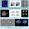
Fig. 2. Emerging imaging measures in Parkinson’s disease.
A Imaging measures for accurate diagnosis of Parkinson’s disease (PD) include: 1. Dopaminergic imaging: axial 123I-FP-CIT single-photon emission computed tomography (SPECT) scans for neuropathologically-confirmed degenerative parkinsonism; adapted from Nicastro et al. (Released under a Creative Commons Attribution-NonCommercial-NoDerivs 4.0 International license). 2. Cardiac meta-iodobenzylguanidine (MIBG) scintigraphy: Absent tracer uptake in the heart (dotted circle) in a patient with dementia with Lewy Bodies (DLB) compared to a control. Heart-to-mediastinum ratios (heart: dotted circles, upper mediastinum: rectangle) can be calculated. Adapted from ref. ; 3. Imaging of the Substantia Nigra (SN): Neuromelanin-sensitive MRI (NM-MRI) to visualise SN atrophy, with earliest changes involving signal loss in nigrosome-1 territory, caudal and mediolateral portions of the SN. Loss of nigrosome-1 shown here in a PD patient in an NM-MRI image (c) and QSM image (d) compared to a control (a: NM-MRI, b: QSM). Adapted with permission from ref. (Released under a Creative Commons Attribution-NonCommercial-NoDerivs 4.0 International license). B Neuroimaging measures for risk stratification and tracking disease progression in PD: 4. Imaging of White Matter: Fixel-based analysis shows reduced white matter fibre cross-section in PD patients who later developed poor outcomes over 3-year follow-up. Colour scale represents percentage reduction in fibre cross-section in PD patients with poor vs PD patients with good outcomes. Adapted from ref. ; 5 Structural connectivity: Widespread reductions in structural connectivity, mostly in frontal and parietal-occipital connections, seen at baseline in PD patients who progress to poor outcomes within 3 years, from ref. ; 6. Free water imaging: Longitudinal changes in free water within the SN seen in PD patients compared to controls. Loss of signal is seen in the midbrain of a PD patient over 4 years. Adapted with permissions, (released under a Creative Commons Attribution-NonCommercial 4.0 International license) 7. Iron-sensitive imaging: QSM signal at baseline correlates with longitudinal worsening of (a)motor and (b)cognitive scores in PD patients over 3 years. Colour bar represents p-value family-wise error-corrected for multiple comparisons, grey scale represents susceptibility values. Adapted from ref. 8. Cortical thickness: Cortical thinning patterns in patients with mild PD compared to controls after 2 and 4-year follow-up. Statistically-significant regions shown in blue, corrected for multiple comparisons using false discovery rate. Colour bar represents t-values. Adapted with permission from Fillippi et al.(Released under a Creative Commons Attribution-NonCommercial-NoDerivs 4.0 International license). CBS Corticobasal syndrome, CTL Control, DLB Dementia with Lewy bodies, L Left; MCI Mild cognitive impairment, MIBG meta-iodobenzylguanidine MSA-P Multiple system atrophy parkinsonian type, NM-MRI Neuromelanin-sensitive MRI, PD Parkinson’s disease, PSP Progressive supranuclear palsy; QSM Quantitative susceptibility mapping, R Right; SN Substantia nigra.

References

    1. Dorsey ER, et al. Global, regional, and national burden of Parkinson’s disease, 1990–2016: a systematic analysis for the Global Burden of Disease Study 2016. Lancet Neurol. 2018;17:939–953. doi: 10.1016/S1474-4422(18)30295-3. - DOI - PMC - PubMed
    1. Irwin DJ, et al. Neuropathological and genetic correlates of survival and dementia onset in synucleinopathies: a retrospective analysis. Lancet Neurol. 2017;16:55–65. doi: 10.1016/S1474-4422(16)30291-5. - DOI - PMC - PubMed
    1. Vijiaratnam N, Simuni T, Bandmann O, Morris HR, Foltynie T. Progress towards therapies for disease modification in Parkinson’s disease. Lancet Neurol. 2021;20:559–572. doi: 10.1016/S1474-4422(21)00061-2. - DOI - PubMed
    1. Simuni, T. et al. Biological Definition of Neuronal alpha-Synuclein Disease: Towards an Integrated Staging System for Research 10.1016/S1474-4422(23)00405-2 (2024). - PubMed
    1. Höglinger GU, et al. A biological classification of Parkinson’s disease: the SynNeurGe research diagnostic criteria. Lancet Neurol. 2024;23:191–204. doi: 10.1016/S1474-4422(23)00404-0. - DOI - PubMed

LinkOut - more resources