

A three-dimensional quantitative assessment on bony growth and symmetrical recovery of mandible after decompression for unicystic ameloblastoma
- PMID: 38969711
- PMCID: PMC11226675
- DOI: 10.1038/s41598-024-66411-4
A three-dimensional quantitative assessment on bony growth and symmetrical recovery of mandible after decompression for unicystic ameloblastoma
Abstract
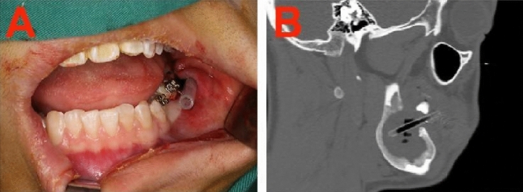
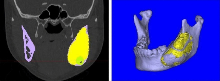
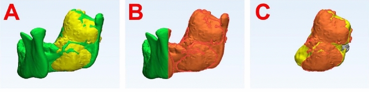
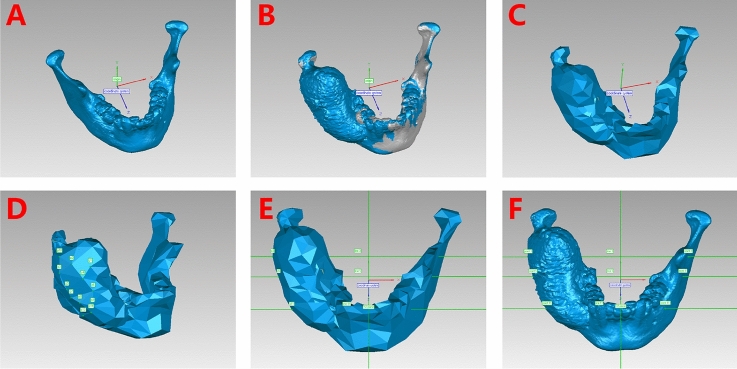
Unicystic ameloblastoma (UAM) of the jaw can be effectively reduced in volume through decompression, which promotes bone regeneration and restores jaw symmetry. This study quantitatively evaluated changes in mandible volume and symmetry following decompression of mandibular UAM. This study included 17 patients who underwent surgical decompression followed by second-stage curettage for mandibular UAM. Preoperative and postoperative three-dimensional computed tomography (CT) images were collected. Bone volume and the area of cortical perforation were measured to assess bone growth during decompression. Mandibular volumetric symmetry was analyzed by calculating the volumetric ratio of the two sides of the mandible. Twelve pairs of landmarks were identified on the surface of the lesion regions, and their coordinates were used to calculate the mean asymmetry index (AI) of the mandible. Paired t-tests and the Mann-Whitney U test were used for statistical analysis, with p < 0.05 considered indicative of statistical significance. The mean duration of decompression was 9.41 ± 3.28 months. The mean bone volume increased by 8.07 ± 2.41%, and cortical perforation recovery was 71.97 ± 14.99%. The volumetric symmetry of the mandible improved significantly (p < 0.05), and a statistically significant decrease in AI was observed (p < 0.05). In conclusion, UAM decompression enhances bone growth and symmetry recovery of the mandible. The present evaluation technique is clinically useful for quantitatively assessing mandibular asymmetry.
Keywords: Ameloblastoma; Bone amount; Cortical perforation; Decompression; Symmetry; Volume reduction.
© 2024. The Author(s).
Conflict of interest statement
The authors declare no competing interests.
Figures
Similar articles
-
To evaluate the clinical efficacy of decompression for large cystic lesions in mandible by digital technology.Eur J Med Res. 2025 Feb 14;30(1):104. doi: 10.1186/s40001-025-02366-0. Eur J Med Res. 2025. PMID: 39953565 Free PMC article.
-
Conservative approach: using decompression procedure for management of a large unicystic ameloblastoma of the mandible.J Craniofac Surg. 2014 May;25(3):1012-4. doi: 10.1097/SCS.0000000000000716. J Craniofac Surg. 2014. PMID: 24699101
-
Marsupialization of mandibular cystic ameloblastoma: Retrospective study of 7 years.Head Neck. 2018 Oct;40(10):2172-2180. doi: 10.1002/hed.25212. Epub 2018 May 13. Head Neck. 2018. PMID: 29756338
-
Unicystic ameloblastoma: a late recurrence with pseudo-glandular features. A case report.Singapore Dent J. 1995 Jul;20(1):21-3. Singapore Dent J. 1995. PMID: 9582685 Review.
-
3D cone beam computed tomography reconstruction images in diagnosis of ameloblastomas of lower jaw: A case report and mini review.J Xray Sci Technol. 2018;26(1):133-140. doi: 10.3233/XST-17344. J Xray Sci Technol. 2018. PMID: 29480235 Review.
Cited by
-
Three-dimensional analysis of natural healing of mandibular bone cavities after cyst enucleation.Clin Oral Investig. 2025 Feb 5;29(2):116. doi: 10.1007/s00784-025-06162-2. Clin Oral Investig. 2025. PMID: 39909920
