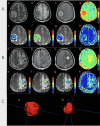
Differentiation of glioma and solitary brain metastasis: a multi-parameter magnetic resonance imaging study using histogram analysis
- PMID: 38969990
- PMCID: PMC11225204
- DOI: 10.1186/s12885-024-12571-5
Differentiation of glioma and solitary brain metastasis: a multi-parameter magnetic resonance imaging study using histogram analysis
Abstract
Background: Differentiation of glioma and solitary brain metastasis (SBM), which requires biopsy or multi-disciplinary diagnosis, remains sophisticated clinically. Histogram analysis of MR diffusion or molecular imaging hasn't been fully investigated for the differentiation and may have the potential to improve it.
Methods: A total of 65 patients with newly diagnosed glioma or metastases were enrolled. All patients underwent DWI, IVIM, and APTW, as well as the T1W, T2W, T2FLAIR, and contrast-enhanced T1W imaging. The histogram features of apparent diffusion coefficient (ADC) from DWI, slow diffusion coefficient (Dslow), perfusion fraction (frac), fast diffusion coefficient (Dfast) from IVIM, and MTRasym@3.5ppm from APTWI were extracted from the tumor parenchyma and compared between glioma and SBM. Parameters with significant differences were analyzed with the logistics regression and receiver operator curves to explore the optimal model and compare the differentiation performance.
Results: Higher ADCkurtosis (P = 0.022), frackurtosis (P<0.001),and fracskewness (P<0.001) were found for glioma, while higher (MTRasym@3.5ppm)10 (P = 0.045), frac10 (P<0.001),frac90 (P = 0.001), fracmean (P<0.001), and fracentropy (P<0.001) were observed for SBM. frackurtosis (OR = 0.431, 95%CI 0.256-0.723, P = 0.002) was independent factor for SBM differentiation. The model combining (MTRasym@3.5ppm)10, frac10, and frackurtosis showed an AUC of 0.857 (sensitivity: 0.857, specificity: 0.750), while the model combined with frac10 and frackurtosis had an AUC of 0.824 (sensitivity: 0.952, specificity: 0.591). There was no statistically significant difference between AUCs from the two models. (Z = -1.14, P = 0.25).
Conclusions: The frac10 and frackurtosis in enhanced tumor region could be used to differentiate glioma and SBM and (MTRasym@3.5ppm)10 helps improving the differentiation specificity.
Keywords: Amide proton transfer-weighted imaging; Glioma; Intravoxel incoherent motion; MRI; Solitary brain metastasis.
© 2024. The Author(s).
Conflict of interest statement
Two authors of this manuscript (J. Guo, and M. Zhang) are the employees of GE Healthcare. No known competing financial interests or personal relationships to declare for the remaining authors.
Figures


Similar articles
-
Bi-exponential diffusion-weighted imaging for differentiating high-grade gliomas from solitary brain metastases: a VOI-based histogram analysis.Sci Rep. 2024 Dec 30;14(1):31909. doi: 10.1038/s41598-024-83452-x. Sci Rep. 2024. PMID: 39738411 Free PMC article.
-
[Value of intravoxel incoherent motion diffusion-weighted imaging in differential diagnosis of benign and malignant hepatic lesions and blood perfusion evaluation].Zhonghua Gan Zang Bing Za Zhi. 2016 Nov 20;24(11):840-845. doi: 10.3760/cma.j.issn.1007-3418.2016.11.009. Zhonghua Gan Zang Bing Za Zhi. 2016. PMID: 27978930 Chinese.
-
Amide proton transfer-weighted imaging to differentiate malignant from benign pulmonary lesions: Comparison with diffusion-weighted imaging and FDG-PET/CT.J Magn Reson Imaging. 2018 Apr;47(4):1013-1021. doi: 10.1002/jmri.25832. Epub 2017 Aug 11. J Magn Reson Imaging. 2018. PMID: 28799280
-
Intravoxel incoherent motion diffusion-weighted imaging analysis of diffusion and microperfusion in grading gliomas and comparison with arterial spin labeling for evaluation of tumor perfusion.J Magn Reson Imaging. 2016 Sep;44(3):620-32. doi: 10.1002/jmri.25191. Epub 2016 Feb 16. J Magn Reson Imaging. 2016. PMID: 26880230
-
Diffusion-Weighted Imaging and Diffusion Tensor Imaging for Differentiating High-Grade Glioma from Solitary Brain Metastasis: A Systematic Review and Meta-Analysis.AJNR Am J Neuroradiol. 2018 Jul;39(7):1208-1214. doi: 10.3174/ajnr.A5650. Epub 2018 May 3. AJNR Am J Neuroradiol. 2018. PMID: 29724766 Free PMC article.
Cited by
-
Sinonasal adenoid cystic carcinoma: preoperative apparent diffusion coefficient histogram analysis in prediction of prognosis and Ki-67 proliferation status.Jpn J Radiol. 2025 Mar;43(3):389-401. doi: 10.1007/s11604-024-01676-3. Epub 2024 Oct 9. Jpn J Radiol. 2025. PMID: 39382794
-
Noninvasive prediction of Ki-67 expression level in IDH-wildtype glioblastoma using MRI histogram analysis: comparison and combination of MRI morphological features.Front Oncol. 2025 May 16;15:1577816. doi: 10.3389/fonc.2025.1577816. eCollection 2025. Front Oncol. 2025. PMID: 40452838 Free PMC article.
References
MeSH terms
LinkOut - more resources
Full Text Sources
Medical

