. 2024 Jul 5;22(1):351.
doi: 10.1186/s12964-024-01728-1.

Tonabersat suppresses priming/activation of the NOD-like receptor protein-3 (NLRP3) inflammasome and decreases renal tubular epithelial-to-macrophage crosstalk in a model of diabetic kidney disease

Affiliations

Tonabersat suppresses priming/activation of the NOD-like receptor protein-3 (NLRP3) inflammasome and decreases renal tubular epithelial-to-macrophage crosstalk in a model of diabetic kidney disease

C L Cliff et al. Cell Commun Signal. .

Abstract

Background: Accompanied by activation of the NOD-like receptor protein 3 (NLRP3) inflammasome, aberrant connexin 43 (Cx43) hemichannel-mediated ATP release is situated upstream of inflammasome assembly and inflammation and contributes to multiple secondary complications of diabetes and associated cardiometabolic comorbidities. Evidence suggests there may be a link between Cx43 hemichannel activity and inflammation in the diabetic kidney. The consequences of blocking tubular Cx43 hemichannel-mediated ATP release in priming/activation of the NLRP3 inflammasome in a model of diabetic kidney disease (DKD) was investigated. We examined downstream markers of inflammation and the proinflammatory and chemoattractant role of the tubular secretome on macrophage recruitment and activation.

Methods: Analysis of human transcriptomic data from the Nephroseq repository correlated gene expression to renal function in DKD. Primary human renal proximal tubule epithelial cells (RPTECs) and monocyte-derived macrophages (MDMs) were cultured in high glucose and inflammatory cytokines as a model of DKD to assess Cx43 hemichannel activity, NLRP3 inflammasome activation and epithelial-to-macrophage paracrine-mediated crosstalk. Tonabersat assessed a role for Cx43 hemichannels.

Results: Transcriptomic analysis from renal biopsies of patients with DKD showed that increased Cx43 and NLRP3 expression correlated with declining glomerular filtration rate (GFR) and increased proteinuria. In vitro, Tonabersat blocked glucose/cytokine-dependant increases in Cx43 hemichannel-mediated ATP release and reduced expression of inflammatory markers and NLRP3 inflammasome activation in RPTECs. We observed a reciprocal relationship in which NLRP3 activity exacerbated increased Cx43 expression and hemichannel-mediated ATP release, events driven by nuclear factor kappa-B (NFκB)-mediated priming and Cx43 hemichannel opening, changes blocked by Tonabersat. Conditioned media (CM) from RPTECs treated with high glucose/cytokines increased expression of inflammatory markers in MDMs, an effect reduced when macrophages were pre-treated with Tonabersat. Co-culture using conditioned media from Tonabersat-treated RPTECs dampened macrophage inflammatory marker expression and reduced macrophage migration.

Conclusion: Using a model of DKD, we report for the first time that high glucose and inflammatory cytokines trigger aberrant Cx43 hemichannel activity, events that instigate NLRP3-induced inflammation in RPTECs and epithelial-to-macrophage crosstalk. Recapitulating observations previously reported in diabetic retinopathy, these data suggest that Cx43 hemichannel blockers (i.e., Tonabersat) may dampen multi-system damage observed in secondary complications of diabetes.

Keywords: Cell-cell communication; Connexin-43; Diabetic nephropathy; Hemichannel; Hyperglycaemia; Inflammasome; Inflammation; Macrophages; NLRP3; Tonabersat.


Conflict of interest statement

The authors declare no competing interests.

Figures

Fig. 1
Fig. 1
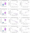
Tonabersat blocks high glucose and pro-inflammatory cytokine-mediated increases in Cx43 hemichannel activity and expression of pro-inflammatory markers in primary human RPTECs. Analysis of Nephroseq transcriptomic datasets show that (a)GJA1 (Cx43) expression increases in kidneys from patients with DN (n = 10 with DN and n = 12 healthy controls) [52] which positively correlates to (b) proteinuria [53] and (c) declining GFR [52]. Primary RPTECs (d) were treated with 5mM/L basal glucose (BG) or 25mM/L high glucose (HG) +/- IL1β (10ng/mL) and TNFα (10ng/mL; Cyt) +/- Tonabersat (TB; 50µM) for 48 h. Carboxyfluorescein dye uptake studies (e) determined change in hemichannel number at the cell surface which was quantified using Fiji software (f). An ATPlite luminescence assay measured cellular release of ATP into the supernatant (g). Use of RT-qPCR evaluated changes in mRNA expression of G-CSF (h), IL1α (i), IL6 (j) and TNFα (k), normalised against GAPDH. All groups represent n = 3–6 unless otherwise specified. ANOVA and Tukey post-test were used for experimental comparisons except for transcriptomic data where an unpaired t-test with Welch’s correction and simple linear regression were used for statistical analysis. Significance is displayed as *P < 0.05 and ***P < 0.001
Fig. 2
Fig. 2
Inflammasome-related gene expression in individuals with diabetic nephropathy correlates to reduced renal function. Transcriptomic analysis was performed on Nephroseq data [52, 53] comparing mRNA expression of IL18 (a & b), IL1β (c), NLRP3 (d-f), caspase 1 (CASP1; g-i) and ASC (j-l) in kidney biopsies from healthy donors and donors with diabetic nephropathy (DN) and compared with functional parameters. The sample number (n) is specified where appropriate. An unpaired t-test with Welch’s correction analysis and simple linear regression were used for statistical analysis. Significance is displayed as *P < 0.05, **P < 0.01, ***P < 0.001
Fig. 3
Fig. 3
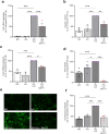
Tonabersat negates high glucose and cytokine-evoked increases in NLRP3 inflammasome priming and activation in human RPTECs. Human RPTECs were treated with 5mM/L basal glucose (BG) or 25mM/L high glucose (HG) +/- IL1β (10ng/mL) and TNFα (10ng/mL; Cyt) +/- Tonabersat (TB; 50µM) for 48 h. RT-qPCR evaluated changes in mRNA expression of IL1β (a) and NLRP3 (b), normalised against GAPDH. Use of a caspase glo-1 assay assessed (c) caspase 1 activity whilst an ELISA assessed changes in IL1β secretion (d). Date representative of n = 3–6 separate experiments. Significance is displayed as *P < 0.05, **P < 0.01, ***P < 0.001
Fig. 4
Fig. 4
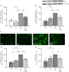
Blocking NFκB-mediated priming and activation of the NLRP3 inflammasome parallels a reduction in Cx43 expression and hemichannel activity. Human RPTECs were treated with 5mM/L basal glucose (BG) or 25mM/L high glucose (HG) +/- IL1β (10ng/mL) and TNFα (10ng/mL; Cyt) +/- BAY11 7082 (5µM) for 48 h. RT-qPCR evaluated changes in IL1β (a) mRNA expression, caspase 1 (b) activity and Cx43 (c) mRNA expression in response to inhibition of NFκB, when normalised against GAPDH. An ATPlite luminescence assay measured cellular release of ATP into the supernatant (d). Carboxyfluorescein dye uptake studies determined changes in hemichannel number at the cell surface (e), quantified using Fiji software (f). Data representative of n = 3–6 separate experiments. Significance is displayed as *P < 0.05, **P < 0.01 ***P < 0.001
Fig. 5
Fig. 5
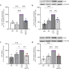
Blocking caspase 1 activity reduces pro-inflammatory cytokine-induced increases in Cx43 expression and hemichannel activity. Human RPTECs were treated with 5mM/L basal glucose (BG) or 25mM/L high glucose (HG) +/- IL1β (10ng/mL) and TNFα (10ng/mL; Cyt) +/- YVAD CMK (10µM) for 48 h. RT-qPCR evaluated changes in Cx43 mRNA expression normalised against GAPDH (a) whilst western blotting (b) assessed altered Cx43 protein expression. Results were normalised against expression of α-Tubulin as a loading control. Carboxyfluorescein dye uptake studies determined change in hemichannel number at the cell surface (c) which was quantified using Fiji software (d). An ATPlite luminescence assay measured cellular release of ATP into the supernatant (e). Data representative of n = 3–6 separate experiments. Significance is displayed as *P < 0.05, **P < 0.01, ***P < 0.001
Fig. 6
Fig. 6
Tonabersat negates a glucose/proinflammatory cytokine-induced increase in Cx43 expression, associated with altered [Ca2+]i gating. Human RPTECs were treated with 5mM/L basal glucose (BG) or 25mM/L high glucose (HG) +/- IL1β (10ng/mL) and TNFα (10ng/mL; Cyt) +/- Tonabersat (TB; 50µM) +/- BAPTA (5µM) for 48 h. RT-qPCR evaluated changes in Cx43 mRNA expression normalised against GAPDH (a) whilst western blotting (b & d) assessed altered Cx43 protein expression. Results were normalised against α-Tubulin as a loading control. An ATPlite luminescence assay measured cellular release of ATP into the supernatant (c). Data is representative of n = 3–5 experiments. Significance is displayed as *P < 0.05, **P < 0.01 and ***P < 0.001
Fig. 7
Fig. 7
Tonabersat reduces tubular epithelial MCP1 expression and resultant paracrine-mediated macrophage migration. Transcriptomic analysis was performed on Nephroseq publicly available data [52, 54] comparing mRNA expression of MCP1 (a), CX3CL1 (g) and CXCL12 (j) in the tubules of kidney biopsies from healthy donors and donors with diabetic nephropathy. The sample number (n) is specified where appropriate. Increased expression of each gene was further correlated to a declining GFR (b, h&k respectively). An unpaired t-test with Welch’s correction analysis and simple linear regression were used for statistical analysis. Human RPTECs were treated with 5mM/L basal glucose (BG) or 25mM/L high glucose (HG) +/- IL1β (10ng/mL) and TNFα (10ng/mL; Cyt) +/- Tonabersat (TB; 50µM) for 48 h. RT-qPCR evaluated changes in MCP1 mRNA expression normalised against GAPDH (d). Conditioned media (CM) generated from treated RPTECs +/- Tonabersat (TB; 50µM) (c) was applied to healthy human monocyte-derived macrophages (MDM) +/- Tonabersat (TB; 50µM) cultured in transwell plate inserts. (e) The number of MDMs migrating into the well containing RPTEC conditioned media was calculated. To support these observations, RT-qPCR evaluated changes in MCP1 (f), CX3CL1 (i) and CXCL12 (l) mRNA expression normalised against GAPDH. Data representative of n = 3–6 experiments. Significance is displayed as *P < 0.05, ** P < 0.01 and ***P < 0.001
Fig. 8
Fig. 8
Blocking Cx43 hemichannels in tubule cells mitigates glucose and inflammatory cytokine-induced paracrine-mediated changes in macrophage polarisation and inflammation. Conditioned media was generated from human RPTECs treated with 5mM/L basal glucose (BG) or 25mM/L high glucose (HG) cultured in IL1β (10ng/mL) and TNFα (10ng/mL; Cyt) +/- Tonabersat (TB; 50µM). This was then applied to healthy human monocyte derived macrophages (MDM) +/- Tonabersat (TB; 50µM) (a). RT-qPCR evaluated changes in expression of both M1 macrophage markers IL1α (b), CD80 (c) and STAT1 (d) and M2 markers IL10 (e), CD206 (f) and STAT6 (g). Data representative of n = 4–6. Significance is displayed as *P < 0.05, **P < 0.01, ***P < 0.001
Fig. 9
Fig. 9
Blocking Cx43 hemichannels in tubule cells and macrophages mitigates glucose and inflammatory cytokine-induced paracrine-mediated changes in macrophage inflammation. Conditioned media was generated from human RPTECs treated with 5mM/L basal glucose (BG) or 25mM/L high glucose (HG) cultured in IL1β (10ng/mL) and TNFα (10ng/mL; Cyt) +/- Tonabersat (TB; 50µM). This was then applied to healthy human monocyte-derived macrophages (MDMs) +/- Tonabersat (TB; 50µM). Changes in mRNA expression of IL1β (a), caspase 1 activity (b) IL6 mRNA (c) and TNFα mRNA expression (d) were assessed. Data representative of n = 5–6. Significance is displayed as *P < 0.05, **P < 0.01, ***P < 0.001
Fig. 10
Fig. 10
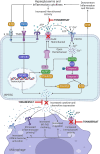
Role of Cx43 hemichannels in induction of injury in a model of diabetic nephropathy via NLRP3 inflammasome priming/activation and paracrine-mediated cell-cell communication. Under hyperglycaemic and pro-inflammatory conditions, proximal tubule epithelial cell Cx43 hemichannels open, releasing ATP. Known to activate purinergic P2 × 7 receptors, ATP leads to K+ efflux and Ca2+ influx, inducing NLRP3 inflammasome complex formation, caspase 1 activation, cleavage of pro-IL1β and release of mature IL1β into the extracellular space. This, in turn, drives signalling pathways culminating in downstream inflammation and fibrosis, including NLRP3 inflammasome priming via translocation of NFκB to the nucleus. Inflammasome priming and activation lead to increased Cx43 expression, elevated [Ca2+]i and increased inflammation, all of which contribute to a further increase in Cx43 hemichannel activity, creating a feedforward loop. As a result of increased inflammatory cytokine and chemokine release, injured RPTECs increased migration, polarisation marker expression and inflammasome priming/activation in donor monocyte-derived macrophages. Hemichannel blocker Tonabersat reduces aberrant Cx43 hemichannel activity in RPTECs and macrophages to negate downstream changes induced by high glucose and inflammation. Created using Biorender.com

Similar articles

Cited by

References

    1. Koye DN, Magliano DJ, Nelson RG, Pavkov ME. The global epidemiology of diabetes and kidney disease. Adv Chronic Kidney Dis. 2018;25:121–32. doi: 10.1053/j.ackd.2017.10.011. - DOI - PMC - PubMed
    1. Cabrera CS, Lee AS, Olsson M, Schnecke V, Westman K, Lind M, et al. Impact of CKD progression on Cardiovascular Disease Risk in a contemporary UK Cohort of individuals with diabetes. Kidney Int Rep. 2020;5:1651. doi: 10.1016/J.EKIR.2020.07.029. - DOI - PMC - PubMed
    1. Bonnet F, Balkau B, Lambert O, Diawara Y, Combe C, Frimat L, et al. The number of nephroprotection targets attained is associated with cardiorenal outcomes and mortality in patients with diabetic kidney disease. The CKD-REIN cohort study. Diabetes Obes Metab. 2024 doi: 10.1111/DOM.15507. - DOI - PubMed
    1. Di Marco M, Scilletta S, Miano N, Marrano N, Natalicchio A, Giorgino F, et al. Cardiovascular risk and renal injury profile in subjects with type 2 diabetes and non-albuminuric diabetic kidney disease. Cardiovasc Diabetol. 2023;22:1–11. doi: 10.1186/S12933-023-02065-2/TABLES/3. - DOI - PMC - PubMed
    1. Bermejo S, González E, López-Revuelta K, Ibernon M, López D, Martín-Gómez A, et al. The coexistence of diabetic retinopathy and diabetic nephropathy is associated with worse kidney outcomes. Clin Kidney J. 2023;16:1656–63. doi: 10.1093/CKJ/SFAD142. - DOI - PMC - PubMed

Publication types

MeSH terms