

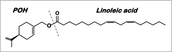
Therapeutic effect of NEO400, perillyl alcohol conjugated to linoleic acid, in a mouse model of UV-induced skin damage
- PMID: 38970228
- PMCID: PMC11913761
- DOI: 10.1111/php.13998
Therapeutic effect of NEO400, perillyl alcohol conjugated to linoleic acid, in a mouse model of UV-induced skin damage
Abstract
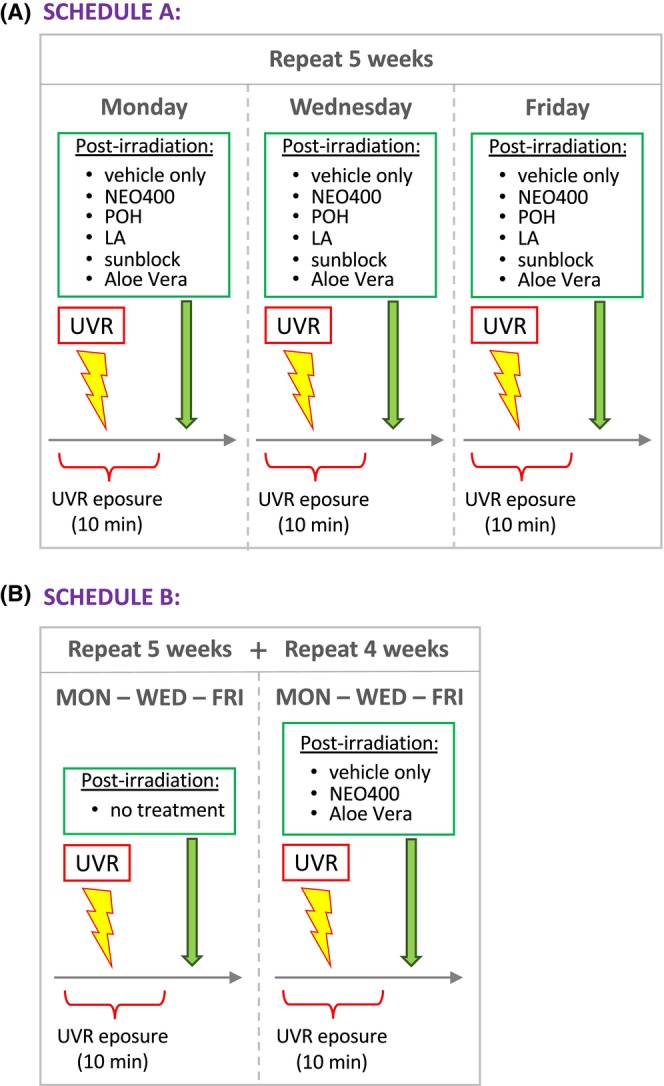
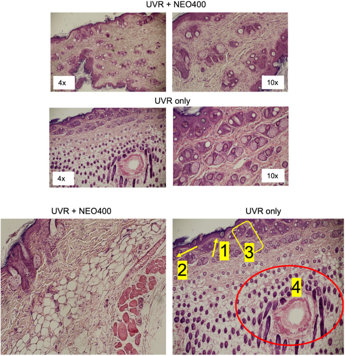
Excessive exposure to ultraviolet radiation (UVR) causes harmful effects on human skin. Pre-exposure application of sunscreen can be protective, but not after damage already has occurred. There is a need for agents that can be applied post-UVR exposure to repair the damage. We investigated a novel compound, NEO400, that appears to meet this medicinal need. NEO400 was created by conjugating linoleic acid to perillyl alcohol. UVR was repeatedly administered to the skin of mice over several weeks, where it caused the typical signs of UV damage, including scaling of the skin, DNA damage, and elevated levels of inflammatory cytokines. However, when NEO400 was applied immediately post-UVR, it triggered the appearance of markers for dermal stem cell proliferation, and no signs of skin damage emerged. Furthermore, when NEO400 was applied to skin that already had incurred significant damage, it accelerated skin healing. When applied individually, linoleic acid and perillyl alcohol were ineffective, indicating that they had to be conjugated in order to exert therapeutic efficacy. None of these skin-protective effects could be achieved with Aloe vera gel, a popular and widely used post-exposure remedy. Our study suggests that NEO400 holds potential as a regenerative treatment for excessively UVR-exposed skin.
Keywords: Aloe vera; UV radiation; linoleic acid; perillyl alcohol; skin damage.
© 2024 The Author(s). Photochemistry and Photobiology published by Wiley Periodicals LLC on behalf of American Society for Photobiology.
Conflict of interest statement
Thomas C. Chen is founder and stakeholder of NeOnc Technologies, Inc. No conflict of interest was declared by the other authors.
Figures


References
-
- Matsumura Y, Ananthaswamy HN. Toxic effects of ultraviolet radiation on the skin. Toxicol Appl Pharmacol. 2004;195:298‐308. - PubMed
-
- Mohania D, Chandel S, Kumar P, et al. Ultraviolet radiations: skin defense‐damage mechanism. Adv Exp Med Biol. 2017;996:71‐87. - PubMed
-
- Garmyn M, Young AR, Miller SA. Mechanisms of and variables affecting UVR photoadaptation in human skin. Photochem Photobiol Sci. 2018;17:1932‐1940. - PubMed
-
- Liang J, Cui L, Li J, Guan S, Zhang K, Li J. Aloe vera: a medicinal plant used in skin wound healing. Tissue Eng Part B Rev. 2021;27:455‐474. - PubMed
MeSH terms
Substances
LinkOut - more resources
Full Text Sources
