

CSF proteomic profiles of neurodegeneration biomarkers in Alzheimer's disease
- PMID: 38970402
- PMCID: PMC11497678
- DOI: 10.1002/alz.14103
CSF proteomic profiles of neurodegeneration biomarkers in Alzheimer's disease
Abstract
Introduction: We aimed to unravel the underlying pathophysiology of the neurodegeneration (N) markers neurogranin (Ng), neurofilament light (NfL), and hippocampal volume (HCV), in Alzheimer's disease (AD) using cerebrospinal fluid (CSF) proteomics.
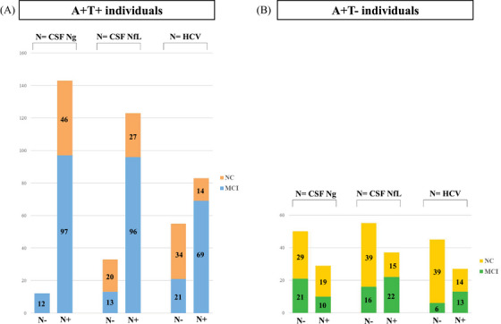
Methods: Individuals without dementia were classified as A+ (CSF amyloid beta [Aβ]42), T+ (CSF phosphorylated tau181), and N+ or N- based on Ng, NfL, or HCV separately. CSF proteomics were generated and compared between groups using analysis of covariance.
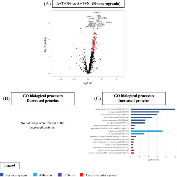
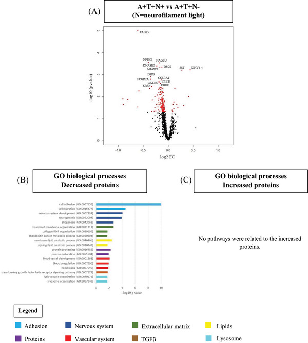
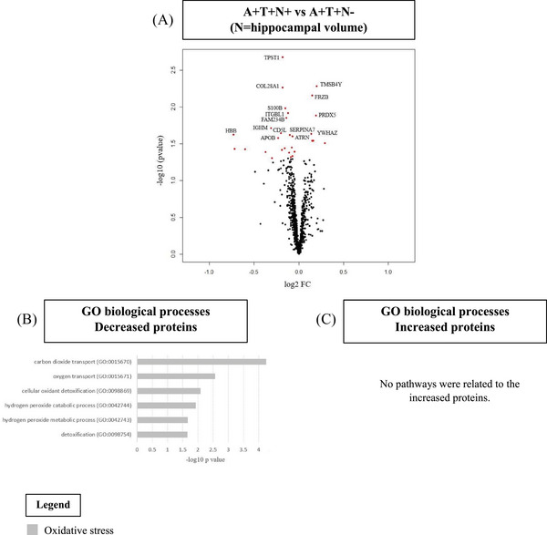
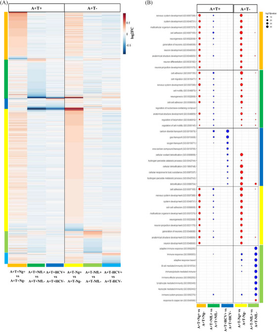
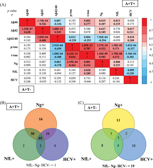
Results: Only a few individuals were A+T+Ng-. A+T+Ng+ and A+T+NfL+ showed different proteomic profiles compared to A+T+Ng- and A+T+NfL-, respectively. Both Ng+ and NfL+ were associated with neuroplasticity, though in opposite directions. Compared to A+T+HCV-, A+T+HCV+ showed few proteomic changes, associated with oxidative stress.
Discussion: Different N markers are associated with distinct neurodegenerative processes and should not be equated. N markers may differentially complement disease staging beyond amyloid and tau. Our findings suggest that Ng may not be an optimal N marker, given its low incongruency with tau pathophysiology.
Highlights: In Alzheimer's disease, neurogranin (Ng)+, neurofilament light (NfL)+, and hippocampal volume (HCV)+ showed differential protein expression in cerebrospinal fluid. Ng+ and NfL+ were associated with neuroplasticity, although in opposite directions. HCV+ showed few proteomic changes, related to oxidative stress. Neurodegeneration (N) markers may differentially refine disease staging beyond amyloid and tau. Ng might not be an optimal N marker, as it relates more closely to tau.
Keywords: Alzheimer's disease; biomarkers; cerebrospinal fluid; hippocampal volume; neurodegeneration markers; neurofilament light; neurogranin; pathophysiology; proteomics.
© 2024 The Author(s). Alzheimer's & Dementia published by Wiley Periodicals LLC on behalf of Alzheimer's Association.
Conflict of interest statement
A.D. received funding from Alzheimer Nederland (grant No. WE.15‐2022‐01). J.G. has nothing to disclose. S.E.S. has analyzed data provided by C2N Diagnostics to Washington University. She has served on scientific advisory boards for Eisai. M.K. has nothing to disclose. L.M.R. has nothing to disclose. V.D. has nothing to disclose. B.M.T. has nothing to disclose. T.L.S.B. has investigator‐initiated research funding from the NIH, the Alzheimer's Association, the Barnes‐Jewish Hospital Foundation, and Siemens. She participates as a site investigator in clinical trials sponsored by Avid Radiopharmaceuticals, Eli Lilly, Biogen, Eisai, Jaansen, and Roche. She serves as a consultant to Biogen, Lilly, Eisai, and Siemens. C.C. has nothing to disclose. C.E.T. has nothing to disclose. I.R. has nothing to disclose. P.M.L. has nothing to disclose. M.T. has nothing to disclose. R.V.’s institution has clinical trial agreements (R.V. as P.I.) with Alector, Biogen, Denali, EliLilly, J&J, UCB. R.V.’s institution has consultancy agreements (R.V. as DSMB member) with AC Immune. J.S. is a senior postdoctoral fellow (12Y1623N) of FWO. J.S. receives funding from Stichting Alzheimer Onderzoek (SAO‐FRA 2021/0022). S.E. has nothing to disclose. E.D.R. has nothing to disclose. J.P. served as a consultant and on advisory boards for the Nestlé Institute of Health Sciences, Ono Pharma, OM Pharma, Schwabe Pharma, Lilly, Roche, and Fujirebio Europe. All his disclosures are unrelated to the present work. The VD cohort was supported by grants from the Swiss National Research Foundation (SNF 320030_204886), Synapsis Foundation – Dementia Research Switzerland (Grant No. 2017‐PI01). G.P. has nothing to disclose. M.T. has nothing to disclose. Y.F.L. has nothing to disclose. S.L. has nothing to disclose. J.S. has nothing to disclose. F.B. is a steering committee or Data Safety Monitoring Board member for Biogen, Merck, Eisai, and Prothena; an advisory board member for Combinostics, Scottish Brain Sciences; a consultant for Roche, Celltrion, Rewind Therapeutics, Merck, Bracco. F.B. has research agreements with ADDI, Merck, Biogen, GE Healthcare, Roche. F.B. is co‐founder and shareholder of Queen Square Analytics LTD. L.B. has nothing to disclose. K.B. has served as a consultant and on advisory boards for AC Immune, Acumen, ALZPath, AriBio, BioArctic, Biogen, Eisai, Lilly, Moleac Pte. Ltd., Novartis, Ono Pharma, Prothena, Roche Diagnostics, and Siemens Healthineers; has served on data monitoring committees for Julius Clinical and Novartis; has given lectures, produced educational materials, and participated in educational programs for AC Immune, Biogen, Celdara Medical, Eisai, and Roche Diagnostics; and is a co‐founder of Brain Biomarker Solutions in Gothenburg AB (BBS), which is a part of the GU Ventures Incubator Program, outside the work presented in this paper. H.Z. has served on scientific advisory boards and/or as a consultant for Abbvie, Acumen, Alector, Alzinova, ALZPath, Amylyx, Annexon, Apellis, Artery Therapeutics, AZTherapies, Cognito Therapeutics, CogRx, Denali, Eisai, Merry Life, Nervgen, Novo Nordisk, Optoceutics, Passage Bio, Pinteon Therapeutics, Prothena, Red Abbey Labs, reMYND, Roche, Samumed, Siemens Healthineers, Triplet Therapeutics, and Wave, has given lectures in symposia sponsored by Alzecure, Biogen, Cellectricon, Fujirebio, Lilly, Novo Nordisk, and Roche, and is a co‐founder of Brain Biomarker Solutions in Gothenburg AB (BBS), which is a part of the GU Ventures Incubator Program (outside submitted work). P.J.V. received funding from the European Commission, IMI 2 Joint Undertaking (JU), AMYPAD, grant No. 115952; European Commission, IMI 2 JU, RADAR‐AD, grant No. 806999; European Commission, IMI 2 JU, EPND, grant No. 101034344. The IMI JU receives support from the European Union's Horizon 2020 research and innovation programme and EFPIA. P.J.V. received also funding from Zon‐MW, Redefining Alzheimer's disease, grant No. 733050824736; and Biogen (Amyloid biomarker study group). Grants were paid to the university. S.J.B.V. received funding from ZonMW (SNAP VIMP grant No. 7330505021), Stichting Adriana van Rinsum‐Ponssen, and the EPND project, which received funding from the European Commision, IMI 2 Joint Undertaking (JU) under grant agreement No. 101034344. The IMI JU receives support from the European Union's Horizon 2020 research and innovation programme and EFPIA. Author disclosures are available in the supporting information.
Figures
References
-
- Wang Z, Yang J, Zhu W, Tang Y, Jia J. The synaptic marker neurogranin as a disease state biomarker in Alzheimer's disease: a systematic review and meta‐analysis. Int J Neurosci. 2021:1‐9. - PubMed
MeSH terms
Substances
Grants and funding
LinkOut - more resources
Full Text Sources
Medical
