. 2024 Jul 6;81(1):287.
doi: 10.1007/s00018-024-05310-3.

Endogenous LXR signaling controls pulmonary surfactant homeostasis and prevents lung inflammation

Affiliations

Endogenous LXR signaling controls pulmonary surfactant homeostasis and prevents lung inflammation

Irene Hernández-Hernández et al. Cell Mol Life Sci. .

Abstract

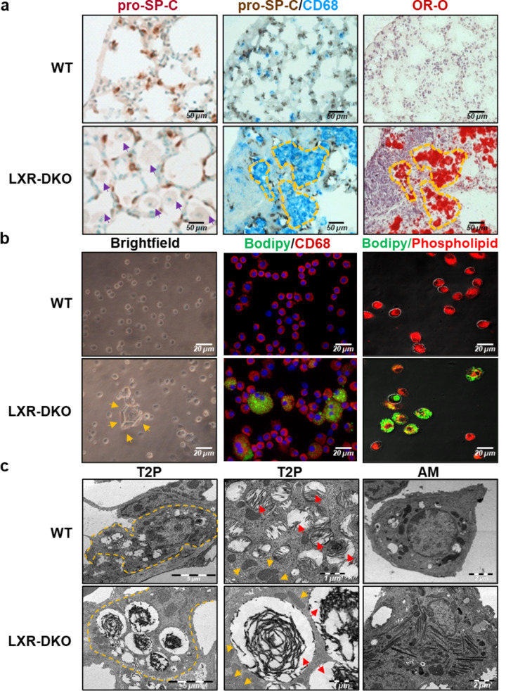
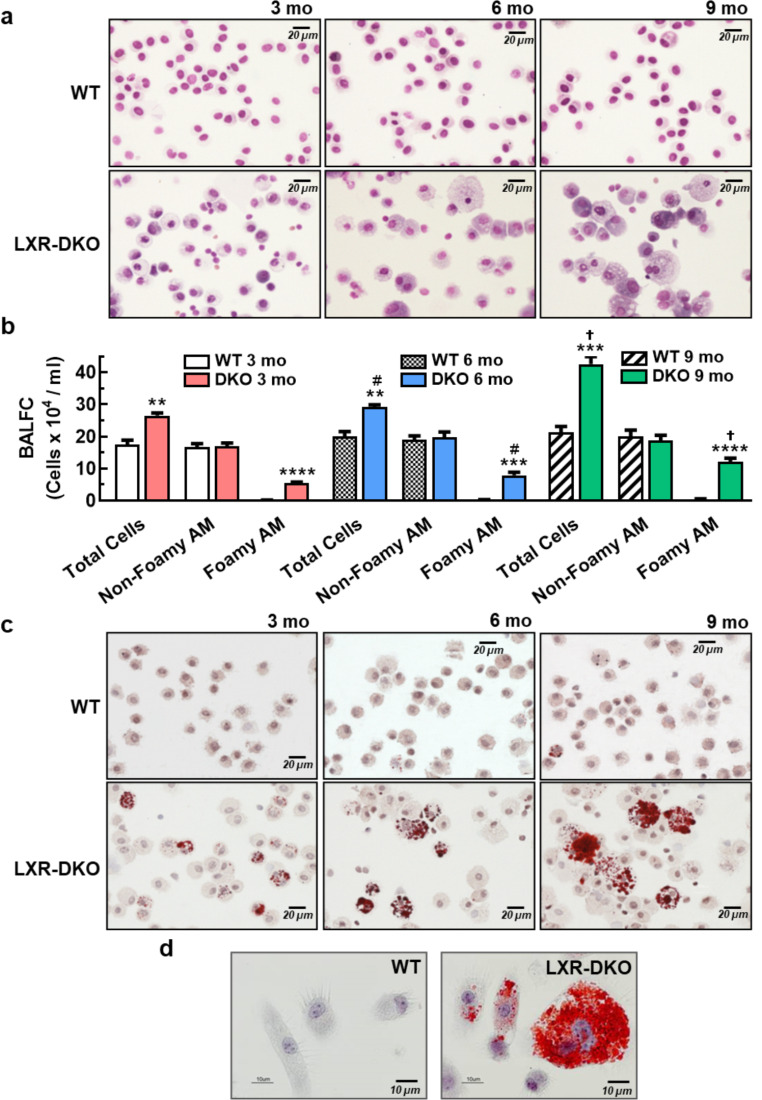
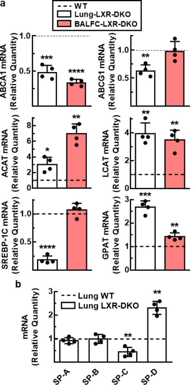
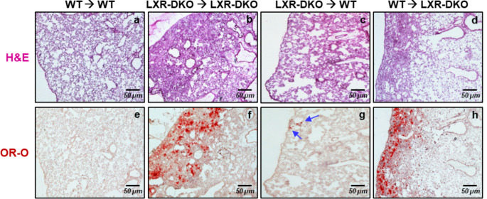
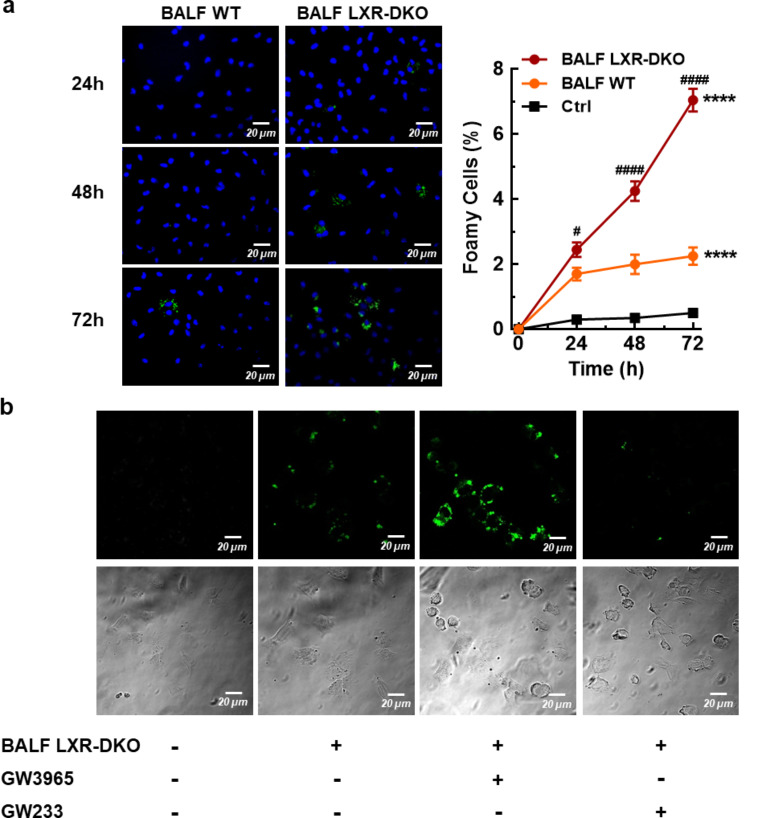
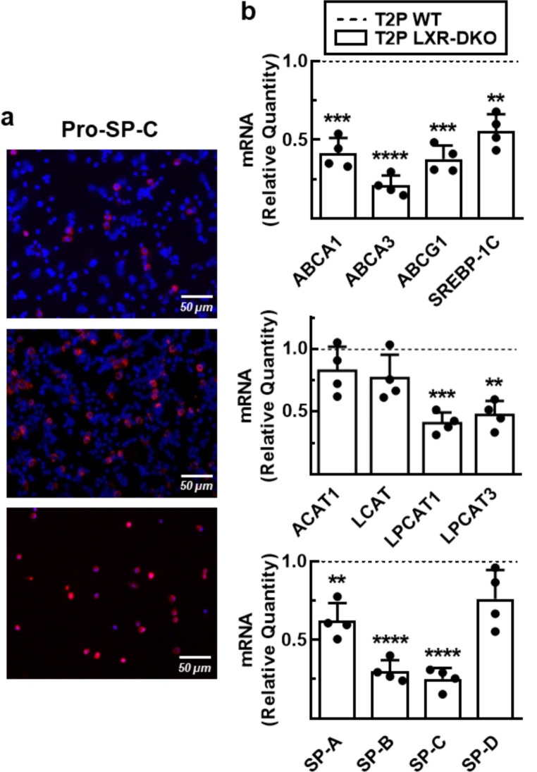
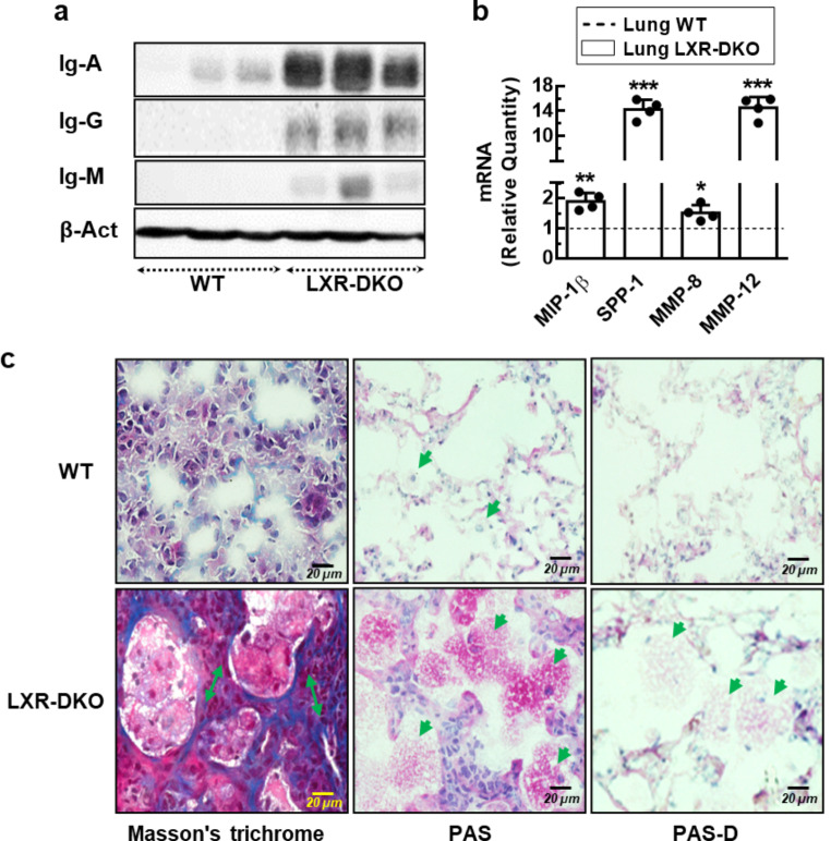
Lung type 2 pneumocytes (T2Ps) and alveolar macrophages (AMs) play crucial roles in the synthesis, recycling and catabolism of surfactant material, a lipid/protein fluid essential for respiratory function. The liver X receptors (LXR), LXRα and LXRβ, are transcription factors important for lipid metabolism and inflammation. While LXR activation exerts anti-inflammatory actions in lung injury caused by lipopolysaccharide (LPS) and other inflammatory stimuli, the full extent of the endogenous LXR transcriptional activity in pulmonary homeostasis is incompletely understood. Here, using mice lacking LXRα and LXRβ as experimental models, we describe how the loss of LXRs causes pulmonary lipidosis, pulmonary congestion, fibrosis and chronic inflammation due to defective de novo synthesis and recycling of surfactant material by T2Ps and defective phagocytosis and degradation of excess surfactant by AMs. LXR-deficient T2Ps display aberrant lamellar bodies and decreased expression of genes encoding for surfactant proteins and enzymes involved in cholesterol, fatty acids, and phospholipid metabolism. Moreover, LXR-deficient lungs accumulate foamy AMs with aberrant expression of cholesterol and phospholipid metabolism genes. Using a house dust mite aeroallergen-induced mouse model of asthma, we show that LXR-deficient mice exhibit a more pronounced airway reactivity to a methacholine challenge and greater pulmonary infiltration, indicating an altered physiology of LXR-deficient lungs. Moreover, pretreatment with LXR agonists ameliorated the airway reactivity in WT mice sensitized to house dust mite extracts, confirming that LXR plays an important role in lung physiology and suggesting that agonist pharmacology could be used to treat inflammatory lung diseases.

Keywords: Alveolar macrophage; Inflammation; LXR; Lipidosis; Surfactant; Type 2 pneumocyte.


Conflict of interest statement

The authors declare that they have no conflict of interest.

Figures

Fig. 1
Fig. 1
Lung histopathology in LXR-deficient mice. a: WT or LXR-DKO lungs from 6-month-old mice were subjected to macroscopic analysis by H&E and oil red O (OR-O) staining, and CD45R/CD3 enzymatic immunohistochemistry. Arrows indicate lipid accumulation (black arrows), tissue congestion (blue arrows), neutral lipid accumulation (yellow arrows), and B- and T-lymphocyte infiltration (green arrows). b: WT or LXR-DKO lungs at 3, 6, 9 and 12 months of age were subjected to OR-O staining. Scale bars of all images are 50 μm. A representative image of 6 mice per genotype is shown
Fig. 2
Fig. 2
Disrupted alveolar physiology in LXR-deficient lungs. a: Consecutive sections of lungs from 6-month-old WT or LXR-DKO mice were subjected to enzymatic immunohistochemistry for the detection of pro-SP-C (left panels), double detection of pro-SP-C and CD-68 (middle panels) or Oil Red O (ORO-O) staining (right panels). Scale bars, 50 μm. Purple arrowheads indicate AMs and areas marked with yellow dashed line indicate coinciding areas with regions occupied by AMs in consecutive sections. A representative image of 5 mice per genotype is shown. b: BAL cells from WT or LXR-DKO 6-month-old mice was subjected to contrast-phase brightfield microscopy (left panels), Bodipy 493/503 fluorescent probe staining (neutral lipids) together with CD68 immunodetection (middle panels), and Bodipy 493/503 together with phospholipid fluorescent probe staining (right panels). Scale bars, 20 μm. Orange arrowheads indicate large hexagonal extracellular crystals. A representative image of 4 mice per genotype is shown. c: Lungs from 6-month-old WT or LXR-DKO mice were subjected to ultrastructural analysis by electron microscopy. Left panels, T2P (8.000 X; Scale bar: 5 μm), areas marked with yellow dashed line indicate cell contours; middle panels, T2P (16.000 X, Scale bar: 1 μm), orange arrowheads indicate mitochondria and red arrowheads indicate lamellar bodies; and right panels, AM (8.000 X, Scale bar: 2 μm). A representative image of 3 mice per genotype is shown
Fig. 3
Fig. 3
Accumulation of lipid-laden AMs in LXR-deficient lungs. a: Representative images (n = 5) of BAL cells, at 3- (left panels), 6- (middle panels) and 9-month-old (right panels) WT and LXR-DKO mice, stained with H&E. Scale bars, 20 μm. b: Total cells, non-foamy and foamy AM in the BAL of 3- to 9-month-old WT and LXR-DKO were counted by contrast-phase brightfield microscopy. Data represent the mean and standard error of three experiments (n = 4 per genotype). Unpaired Student’s t test was used for two-group comparisons; **p < 0.01; ***p < 0.001; ****p < 0.0001 compared to WT group; #p < 0.05 compared to 3 months old and P < 0.05 compared to 6 months old mice. c: BAL cells from3- (left panels), 6- (middle panels) and 9-month-old (right panels) WT and LXR-DKO mice were subjected to Oil-Red-O staining and analyzed by contrast-phase brightfield microscopy. Scale bars, 20 μm. d, Representative images of BAL cells from 9-month-old WT and LXR-DKO mice (n = 4 per genotype) stained with Oil-Red-O (100x). Scale bars, 10 μm
Fig. 4
Fig. 4
Impaired pulmonary surfactant homeostasis gene expression in LXR-deficient mice. Transcript levels of genes involved in lipid (a) and protein (b) metabolism of pulmonary surfactant from BAL cells (BALC) and lung tissue from WT and LXR-DKO 6-month-old mice were analyzed by real-time qPCR. WT gene expression values were normalized to 1 and used as control (dashed line). Data represent the mean and standard error of three experiments (n = 4 mice per genotype). Unpaired Student’s t test with Welch’s correction was used for two-group comparisons; *p < 0.05; **p < 0.01; ***p < 0.001; ****p < 0.0001 compared to WT group
Fig. 5
Fig. 5
Lung histopathology and lipid accumulation in WT and LXR-DKO mice after bone marrow transplant. Morphology (H&E) and lipidosis (Oil-Red-O staining) were assessed in consecutive lung sections from wild-type and LXR-DKO mice 16 weeks after reciprocal bone marrow transplantation (labels: donor→recipient). Scale bars, 50 μm. Images are representative of two independent experiments with five to six mice per group
Fig. 6
Fig. 6
Modulation of LXR activity affects the handling of surfactant material by MLE12 cells. In vitro lipidosis model: Peritoneal macrophages from WT mice were cultured and exposed to BALF from WT and LXR-DKO mice. Subsequently, (a) fluorescence microscopy analysis and percentage of foam cells were determined using Bodipy 493/503 after 24, 48 and 72 h incubation of peritoneal macrophages with BALF. A representative image is shown (left panel) and the data are presented as the mean and standard error (right panel), n = 4 mice per genotype. Kruskal-Wallis with a post hoc Dunn’s test was used for multiple comparisons; ****p < 0.0001 and **p < 0.001 compared to control group; #P < 0.05 and ##P < 0.001 compared to BALF WT groups. (b) MLE-12 cells were pre-incubated with GW3965 agonist or GW233 antagonist for 24 h and then exposed to LXR-DKO BALF for additional 24 h. Lipid uptake was measured by bodipy 493/503 signal (A-G) or bright field (B-H) using confocal microscopy. Cells not exposed to BALF were used as control. A representative image of 3 independent experiments is shown. Scale bars of all images are 50 μm
Fig. 7
Fig. 7
LXR deficiency alters primary type 2 pneumocytes gene expression. a, T2Ps were isolated from WT and LXR-DKO 6-month-old mice by enzymatic digestion and immunomagnetic purification, and subsequently cultured on a Matrigel-coated surface. Enrichment of T2P through the isolation method was monitored by pro-SPC expression by immunocytochemistry in the total lung homogenate (upper panel), after recovery of non-adherent cells (middle panel) and at the end point of isolation after the positive selection column (low panel). A representative image (n = 4) is shown. Scale bars of all images are 50 μm. b, Transcript levels of genes involved in lipid (upper and middle panels) and protein (low panel) metabolism of pulmonary surfactant from T2P, isolated from WT and LXR-DKO 6-month-old mice, were analyzed by real-time qPCR. WT gene expression values were normalized to 1 and used as control (dashed line). Data represent the mean and standard error of three experiments (n = 4 mice per genotype). Unpaired Student’s t test with Welch’s correction was used for two-group comparisons; **p < 0.01; ***p < 0.001; ****p < 0.0001 compared to the WT T2P group
Fig. 8
Fig. 8
LXR inactivation leads to a chronic inflammatory process in the lung. a, Whole cell lysates were prepared from homogenized lung tissue cells from perfused WT and LXR-DKO 6-month-old mice and IgA, IgM or IgG proteins were analyzed by Western blot. Membranes were stripped and reprobed with β−actin antibody as loading control. A representative western blot (n = 3 mice) is shown. b, Transcript levels of inflammatory related-genes, from homogenized lung tissue cells from WT and LXR-DKO 6-month-old mice, were analyzed by real-time qPCR. WT gene expression values were normalized to 1 and used as control (dashed line). Data represent the mean and standard error of three experiments (n = 3 mice per genotype). Unpaired Student’s t test with Welch’s correction was used for two-group comparisons; *p < 0.05; **p < 0.01; ***p < 0.001 compared to WT group. c, Masson’s trichrome (MT) staining (left panels) and periodic acid-Schiff (PAS) staining (middle panels) were evaluated in consecutive lung sections from 6-month-old WT and LXR-DKO mice. Pretreatment of tissues with PAS-diastase (α-salivary amylase) (PAS-D) (right panels) was assessed to confirm PAS-positive material. A representative image of 4 mice per genotype is shown. Scale bars of all images are 20 μm
Fig. 9
Fig. 9
LXR pharmacological activation ameliorates lung reactivity in mice exposed to HDM. WT or LXR-DKO 3-month-old mice were administered with HDM extract daily intranasally at a dose of 25 µg/mouse for 10 consecutive days. a, Lung resistance (RL) to increasing doses of methacholine was assessed 24 h after the last HDM exposure. Data represent the mean and standard error of n = 5 (WT) or n = 4 (LXR-DKO) mice. The effect of genotype was analyzed using one-way ANOVA with a post hoc Bonferroni test; * p < 0.05. b, Histological analysis of the number (left panel) of perivascular and/or peribronchiolar inflammatory infiltrates in the lung. The lung inflammation score (right panel) was calculated as described in Methods. Unpaired Student’s t test; *p < 0.05, **p < 0.01 compared to WT group. c, Representative images from the lungs of HDM-exposed WT or LXR-DKO mice. d, 8- week- old WT mice were separated in three groups: non-sensitized mice (Saline group, N = 11) received daily an intranasal administration of physiological saline and an intraperitoneal administration of vehicle (DMSO in PBS); HDM-sensitized mice received daily an intranasal administration of HDM and an intraperitoneal administration of either vehicle (DMSO in PBS) (HDM group, N = 10) or of the LXR agonist GW3965 (20 mg/kg) (GW3965 + HDM group, N = 11). Administration of the LXR agonist or vehicle was initiated one day before the start of sensitization. Lung resistance (RL) to increasing doses of methacholine was assessed 24 h after the last HDM exposure. Pooled data from two independent experiments. The effect of treatment was analyzed using one-way ANOVA with a post hoc Bonferroni test. Mean ± SEM; ###P < 0.0001 compared to saline group; *p < 0.01 compared to HDM group

References

    1. Cho SJ, Stout-Delgado HW. Aging and lung disease. Annu Rev Physiol. 2020;82:433–459. doi: 10.1146/annurev-physiol-021119-034610. - DOI - PMC - PubMed
    1. Nakamura A, Ebina-Shibuya R, Itoh-Nakadai A, et al. Transcription repressor Bach2 is required for pulmonary surfactant homeostasis and alveolar macrophage function. J Exp Med. 2013;210:2191–2204. doi: 10.1084/jem.20130028. - DOI - PMC - PubMed
    1. Fehrenbach H. Alveolar epithelial type II cell: defender of the alveolus revisited. Respir Res. 2001;2:33. doi: 10.1186/rr36. - DOI - PMC - PubMed
    1. Castranova V, Rabovsky J, Tucker JH, Miles PR. The alveolar type II epithelial cell: a multifunctional pneumocyte. Toxicol Appl Pharmacol. 1988;93:472–483. doi: 10.1016/0041-008x(88)90051-8. - DOI - PubMed
    1. Bissonnette EY, Lauzon-Joset J-F, Debley JS, Ziegler SF. Cross-talk between Alveolar macrophages and Lung epithelial cells is essential to maintain lung homeostasis. Front Immunol. 2020;11:583042. doi: 10.3389/fimmu.2020.583042. - DOI - PMC - PubMed

MeSH terms

LinkOut - more resources