. 2024 Jul;10(4):e12389.
doi: 10.1002/2056-4538.12389.

Mesonephric-type adenocarcinomas of the ovary: prevalence, diagnostic reproducibility, outcome, and value of PAX2

Affiliations

Mesonephric-type adenocarcinomas of the ovary: prevalence, diagnostic reproducibility, outcome, and value of PAX2

Martin Köbel et al. J Pathol Clin Res. 2024 Jul.

Abstract

Mesonephric-type (or -like) adenocarcinomas (MAs) of the ovary are an uncommon and aggressive histotype. They appear to arise through transdifferentiation from Müllerian lesions creating diagnostic challenges. Thus, we aimed to develop a histologic and immunohistochemical (IHC) approach to optimize the identification of MA over its histologic mimics, such as ovarian endometrioid carcinoma (EC). First, we screened 1,537 ovarian epithelial neoplasms with a four-marker IHC panel of GATA3, TTF1, ER, and PR followed by a morphological review of EC to identify MA in retrospective cohorts. Interobserver reproducibility for the distinction of MA versus EC was assessed in 66 cases initially without and subsequently with IHC information (four-marker panel). Expression of PAX2, CD10, and calretinin was evaluated separately, and survival analyses were performed. We identified 23 MAs from which 22 were among 385 cases initially reported as EC (5.7%) and 1 as clear cell carcinoma. The interobserver reproducibility increased from fair to substantial (κ = 0.376-0.727) with the integration of the four-marker IHC panel. PAX2 was the single most sensitive and specific marker to distinguish MA from EC and could be used as a first-line marker together with ER/PR and GATA3/TTF1. Patients with MA had significantly increased risk of earlier death from disease (hazard ratio = 3.08; 95% CI, 1.62-5.85; p < 0.0001) compared with patients with EC, when adjusted for age, stage, and p53 status. A diagnosis of MA has prognostic implications for stage I disease, and due to the subtlety of morphological features in some tumors, a low threshold for ancillary testing is recommended.

Keywords: GATA3; PAX2; TTF1; mesonephric; mesonephric‐like.


Figures

Figure 1
Figure 1
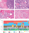
Examples of classic mesonephric‐type adenocarcinoma (MA) with a blue low‐power appearance in a pink collagenous background in (A) and (C) with an admixture of architectural patterns including (A) dense tubules and sieve‐like and (B) papillary and tubular. In corresponding higher power images (C and D), eosinophilic luminal material is conspicuous. There is nuclear overlapping in (C) and open chromatin, grooves, and angulated contours/indentations are apparent in (D). (E) Heat map showing biomarker characteristics of the MA cases as well as presence of associated endometriosis. The detailed ER/PR expression reported as % distribution and (three‐tier intensity) for the five positive MAs were as follows: MA#29 ER 100%(3)/PR 1%(1); MA#24 ER 40%(1)/PR 1%(2); MA#25 ER 5%(1)/PR 20%(3); MA#26 ER 0%/PR 10%(2); MA#27 ER 0%/PR 5%(2).
Figure 2
Figure 2
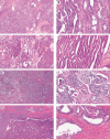
Cases from the reproducibility set with persistent issues where immunohistochemistry was not ordered. (A and B) MA#02 with angulated ducts and cribriform architecture. Eosinophilic luminal material is absent, and the cell shape is more cuboidal. (C and D) MA#03 with more typical long tubular and papillary architectures and cuboidal cell shape. (E and F) MA#04 with nested and spindled architecture and focal eosinophilic luminal material. (G and H) MA#05 with nested/solid architecture and angulated ducts with focal eosinophilic luminal material. Some columnar cells with apical secretions and focal cilia are noted but the predominant cell shape is cuboidal.
Figure 3
Figure 3
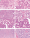
Cases from the reproducibility set with persistent issues of immunohistochemistry (IHC) integration including cases that did not show a mesonephric‐type IHC profile. (A and B) MA#12 with nested/solid architecture and angulated ducts as well as focal eosinophilic luminal material; double‐negative IHC profile but MA methylation profile and normal retained PAX2 expression. (C and D) MA#28 with nested/solid architecture and angulated ducts with focal eosinophilic luminal material; double‐negative IHC profile but MA methylation profile and normal retained PAX2 expression. (E and F) MA#29 with papillary and angulated ductal architecture and cuboidal cell shape and conspicuous eosinophilic luminal material, double‐positive IHC profile (diffuse ER expression) and MA methylation profile as well as normal retained PAX2 expression. (G and H) MA#18 with pale low‐power appearance, angulated ductal architecture, cuboidal cell shape, eosinophilic luminal material and typical nuclear features. The case had a MA‐IHC profile with only focal GATA3 expression.
Figure 4
Figure 4
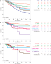
Kaplan–Meier survival analysis; p value – log‐rank test. (A) Comparison of mesonephric‐type adenocarcinomas (MA) to other histotypes [low and high grade serous, endometrioid (EC), clear cell, and mucinous carcinomas]. (B) Grouped immunohistochemical (IHC) profiles of EC (initial diagnosis). (C) Comparison of MA to grouped IHC profiles of EC (diagnosis after MA reclassification). Tables on the right indicate numbers at risk.
Figure 5
Figure 5
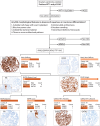
Hierarchical decision tree using combined morphologic and immunohistochemistry (IHC)‐based identification of mesonephric‐type adenocarcinoma (MA). First, a combination of WT1+ and p53 abnormal is highly specific for high‐grade serous carcinoma (HGSC). Second, there are typical morphological features of MA; however, they can overlap with endometrioid carcinomas (EC). Third, to distinguish EC from MA, p53abn and MMRd molecular subtypes of EC are generally inconsistent with MA. Fourth, the most efficient approach to distinguish MA from EC, NSMP molecular subtype (alternative approach without PAX2 is shown in supplementary material, Figure S5).

References

    1. McCluggage WG. Mesonephric‐like adenocarcinoma of the female genital tract: from morphologic observations to a well‐characterized carcinoma with aggressive clinical behavior. Adv Anat Pathol 2022; 29: 208–216. - PubMed
    1. McFarland M, Quick CM, McCluggage WG. Hormone receptor‐negative, thyroid transcription factor 1‐positive uterine and ovarian adenocarcinomas: report of a series of mesonephric‐like adenocarcinomas. Histopathology 2016; 68: 1013–1020. - PubMed
    1. WHO Classification of Tumours Editorial Board . Female Genital Tumours (5th edn). International Agency for Research on Cancer: Lyon, 2020.
    1. McCluggage WG, Vosmikova H, Laco J. Ovarian combined low‐grade serous and mesonephric‐like adenocarcinoma: further evidence for a Mullerian origin of mesonephric‐like adenocarcinoma. Int J Gynecol Pathol 2020; 39: 84–92. - PubMed
    1. da Silva EM, Fix DJ, Sebastiao APM, et al. Mesonephric and mesonephric‐like carcinomas of the female genital tract: molecular characterization including cases with mixed histology and matched metastases. Mod Pathol 2021; 34: 1570–1587. - PMC - PubMed

MeSH terms