. 2024 Jul 6;15(1):5680.
doi: 10.1038/s41467-024-50077-7.

ATP6V0A1-dependent cholesterol absorption in colorectal cancer cells triggers immunosuppressive signaling to inactivate memory CD8+ T cells

Affiliations

ATP6V0A1-dependent cholesterol absorption in colorectal cancer cells triggers immunosuppressive signaling to inactivate memory CD8+ T cells

Tu-Xiong Huang et al. Nat Commun. .

Abstract

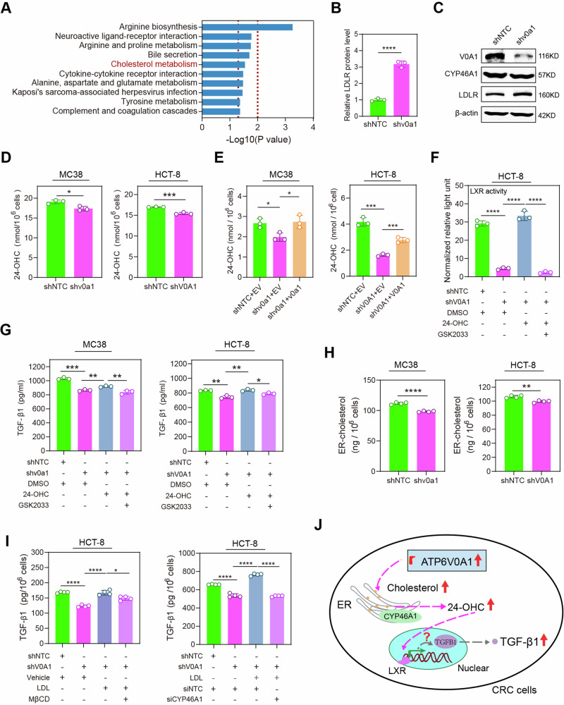
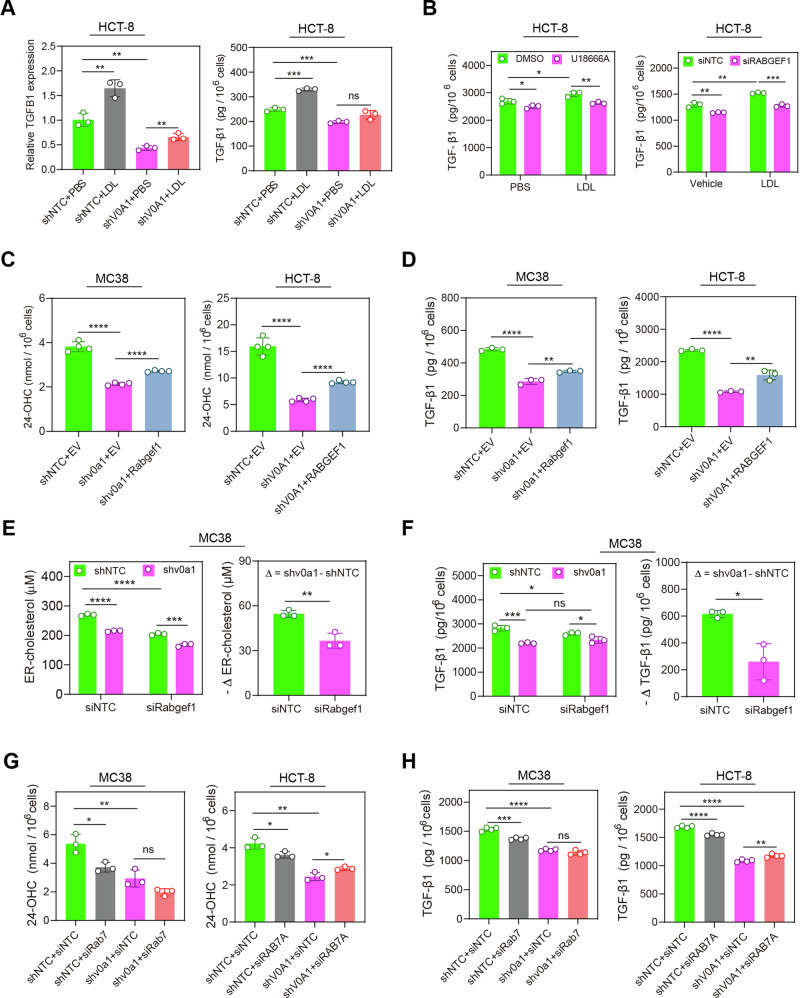
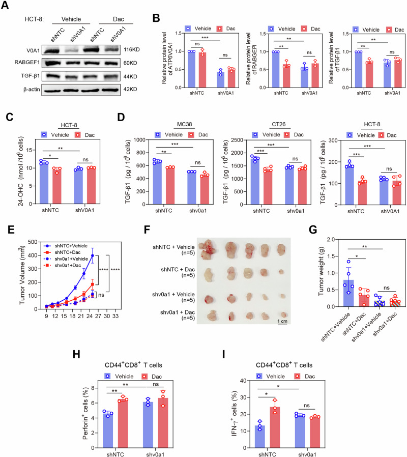
Obesity shapes anti-tumor immunity through lipid metabolism; however, the mechanisms underlying how colorectal cancer (CRC) cells utilize lipids to suppress anti-tumor immunity remain unclear. Here, we show that tumor cell-intrinsic ATP6V0A1 drives exogenous cholesterol-induced immunosuppression in CRC. ATP6V0A1 facilitates cholesterol absorption in CRC cells through RAB guanine nucleotide exchange factor 1 (RABGEF1)-dependent endosome maturation, leading to cholesterol accumulation within the endoplasmic reticulum and elevated production of 24-hydroxycholesterol (24-OHC). ATP6V0A1-induced 24-OHC upregulates TGF-β1 by activating the liver X receptor (LXR) signaling. Subsequently, the release of TGF-β1 into the tumor microenvironment by CRC cells activates the SMAD3 pathway in memory CD8+ T cells, ultimately suppressing their anti-tumor activities. Moreover, we identify daclatasvir, a clinically used anti-hepatitis C virus (HCV) drug, as an ATP6V0A1 inhibitor that can effectively enhance the memory CD8+ T cell activity and suppress tumor growth in CRC. These findings shed light on the potential for ATP6V0A1-targeted immunotherapy in CRC.


Conflict of interest statement

The authors declare no competing interests.

Figures

Fig. 1
Fig. 1. ATP6V0A1 is required for HFD-induced suppression of anti-tumor immunity.
A Schematic diagram showing an animal model to investigate the significance of CRC cell-derived ATP6V0A1 in the suppression of anti-tumor immunity induced by elevated level of exogenous lipids. BD C57BL/6 J mice were fed with high-fat diet (HFD) or control diet (CD) for 9 weeks prior to tumor cell implantation, and the body weight was measured (B); using a body-weight randomization grouping approach, HFD mice and CD mice were separately divided into two groups with similar body weights (C) and comparable serum levels of LDL-cholesterol (D). EH Control MC38 (shNTC) cells or ATP6v0a1-knockdown (shv0a1) MC38 cells were subcutaneously injected to HFD mice and CD mice as shown in (A). Tumor volumes were monitored using calipers, and average tumor growth curves were plotted (E); photographs of the tumors are shown in (F). Tumor-infiltrating lymphocytes (TILs) were isolated and co-cultured with CFSE-labeled MC38 cells, and the cell mixture was analyzed by flow cytometry for the proportion of tumor cell death to assess the killing activities of TILs (G). The relative cytotoxicity of TILs between CD-treated and HFD-treated MC38-shNTC tumors or between CD-treated and HFD-treated MC38-shv0a1 tumors (H) were assessed by calculating the ratio of tumor cell-death proportion (G) between the cell mixture containing TILs from these tumors. For all experiments, data are shown as means ± s.e.m; *p < 0.05, **p < 0.01, ***p < 0.001, ****p < 0.0001. Statistical significance was determined using ordinary two-way ANOVA (in B, E) or unpaired two-sided Student’s t-test (in C, D, G, H). n = 10 (B), 5 (CF), or 3 (G, H) mice in each group; Data representative three independent experiments (BH). Source data and exact p-value are provided as a Source Data file.
Fig. 2
Fig. 2. ATP6V0A1 promotes the growth of colorectal cancers in an immune-dependent manner.
A The effect of suppressing or overexpressing Atp6v0a1 on the proliferation rates of MC38 cells in vitro was analyzed by CCK8 assay. n = 3 independent experiments. BD C57BL/6 J mice were subcutaneously injected with Atp6v0a1-suppressing or Atp6v0a1-overexpressing MC38 cells as shown in Supplementary Fig. 6A. Tumor volumes were monitored using calipers, and average tumor growth curves were plotted (B); photographs of the tumors are shown in (C). n = 5 mice per group. TILs isolated from the tumors described in (B) and (C) were co-cultured with CFSE-labeled MC38 cells, and the cell mixture was analyzed by flow cytometry for the proportion of tumor cell death to assess the killing activities of TILs (D). n = 3 mice in each group; Data representative three independent experiments. EH Atp6v0a1-suppressing or Atp6v0a1-overexpressing MC38 cells were subcutaneously injected into C57BL/6 Rag2−/−Il2rg−/− mice (E, G) or NOD/SCID mice (F, H) as shown in supplementary Fig. 6A. Average tumor growth curves were plotted (E, F); photographs of the tumors are shown in (G, H). n = 4 (E, G) or 5 (F, H) mice per group. I The proliferation of control HCT-8 (shNTC) cells and ATP6V0A1-knockdown (shV0A1) HCT-8 cells in vitro was analyzed by CCK8 assay. n = 3 independent experiments. J, K HCT-8 shNTC cells or HCT-8 shV0A1 cells were subcutaneously injected into huPBMC-NCG mice and control NCG mice as shown in Supplementary Fig. 6B. Average tumor growth curves were plotted (J); photographs of the tumors are shown in (K). n = 4 (shNTC group in huPBMC-NCG mice model) or 5 mice (other groups) in each group. LO Cecal MC38 tumors were established as illustrated in (L). Tumor growth was monitored via bioluminescence detection, and average tumor growth curves were plotted (n = 5 mice per group; M). The mice were sacrificed on day 25; photographs of the tumors in the cecum (N, upper) and the isolated tumors (N, lower) are shown. Tumor weights for the indicated groups are compared in (O). For all experiments, data are shown as means ± s.e.m; *p < 0.05, **p < 0.01, ***p < 0.001, ****p < 0.0001. Statistical significance was determined using ordinary two-way ANOVA (A, B, E, F, I, J, M) or unpaired two-sided Student’s t-test (D, O). Source data and exact p-value are provided as a Source Data file.
Fig. 3
Fig. 3. Memory CD8+ T cell effectiveness is important for the regulation of CRC growth by tumor-derived ATP6V0A1.
AC CD8+ T cells were depleted from C57BL/6 mice in the indicated groups by injection of anti-CD8α mAb (clone 2.43) at the time points shown (A). Average tumor growth curves were plotted (B); photographs of the tumors are shown in (C). n = 5 mice per group. D Flow cytometry (FC) strategy for gating CD44+CD8+ and CD44-CD8+ T cells and detecting effector cytokines. E BCL2 protein levels were compared in CD44+CD8+ and CD44CD8+ T cells by FC. n = 6; Data are pooled from 2 independent experiments. FI The effectiveness of tumor-infiltrating CD44+CD8+ and CD44CD8+ T cells or CD45RO+CD8+ and CD45ROCD8+ T cells was analyzed via FC in subcutaneous MC38 tumors from C57BL/6 J mice (F), subcutaneous CT26 tumors from BALB/c mice (G), subcutaneous HCT-8 tumors from huPBMC-NCG mice (H), and cecal MC38 tumors from C57BL/6 J mice (I). n = 3 mice per group; Data representative of 3 independent experiments. For all experiments, data are shown as means ± s.e.m; *p < 0.05, **p < 0.01, ***p < 0.001. Statistical significance was determined using ordinary two-way ANOVA (B) or unpaired two-sided Student’s t-test (EI). Source data and exact p-value are provided as a Source Data file.
Fig. 4
Fig. 4. Tumor cell-intrinsic ATP6V0A1 suppresses memory CD8+ T cells via the TGF-β1/SMAD3 axis.
A Atp6v0a1 knockdown-induced expression changes in Smad3 and Id2 were analyzed in the indicated T-cell subpopulations based on the scRNA-seq data (Supplementary Fig. 8). B Western blotting detecting the indicated protein levels in CD8+ T cells isolated from MC38-shNTC and MC38-shv0a1 tumors. The samples derive from the same experiment but different gels for SMAD3, ID2, GAPDH, another for p-SMAD3 were processed in parallel. C scRNA-seq data comparing the expression of Tgfbr2 in different T-cell subpopulations. D FC-analysis comparing TGF-βRII level between CD44+CD8+ and CD44-CD8+ T cells in wild-type MC38 tumors. E, F The mRNA and protein levels of TGF-β1 in MC38 cells were analyzed by qPCR (E, left), Western blotting (E, medium), and ELISA (E, right and F). GI Wild-type MC38 tumor-derived CD8+ T cells were treated with the following: (E)-SIS3 (G), MC38 cell culture medium plus control IgG or anti-TGF-β1 (H), conditioned medium from MC38-shNTC cells or MC38-shv0a1 cells with/without supplement of TGF-β1 protein (I). The activation of CD44+ CD8+ and CD44-CD8+ T cells was determined by FC analysis. JL MC38-EV, or MC38-v0a1 cells were subcutaneously injected into C57BL/6 J mice, followed by intraperitoneal injection of control IgG or anti-TGF-β1 on days 7, 10, and 13. Average tumor growth curves (J), photographs of the tumors (K), and a comparison of tumor weights on day 18 (L) are shown; n = 5 mice per group. MO MC38-shNTC or MC38-shv0a1 cells were subcutaneously injected into C57BL/6 J mice; PBS or mouse TGF-β1 protein was injected intratumorally. Average tumor growth curves (M), photographs of the tumors (N), and a comparison of tumor weights on day 20 (O, left) are shown. TILs from day 20 tumors were analyzed for effector production in CD44+CD8+ T cells (O, right). n = 4 mice per group. For all experiments, data are shown as means ± s.e.m; *p < 0.05, **p < 0.01, ***p < 0.001, ****p < 0.0001. Statistical significance was determined using ordinary two-way ANOVA (in J, M) or unpaired two-sided Student’s t-test (in D, E, F, G, H, I, L, O). n = 3 independent experiments for Fig. 4D–I; Three independent experiments were performed for Fig. 4B, E (medium). Source data and exact p-value are provided as a Source Data file.
Fig. 5
Fig. 5. Cholesterol accumulation in the ER is essential for the upregulation of TGF-β1 by ATP6V0A1 in CRC cells.
A, B Atp6v0a1 knockdown-induced changes in protein levels in MC38 cells were analyzed using label-free protein quantitative mass spectrometry (MS); KEGG route enrichment analysis showed that changes in the cholesterol metabolism pathway were induced by Atp6v0a1 knockdown (A). Relative levels of LDLR protein in MC38-shNTC and MC38-shv0a1 cells were determined by MS analysis (B; n = 3 independent experiments). C Western blotting was used to detect LDLR and CYP46A protein levels in MC38-shNTC and MC38-shv0a1 cells. The samples derive from the same experiment but different gels for ATP6V0A1, CYP46A1, β-actin, another for LDLR were processed in parallel. Data representative of 3 independent experiments. D, E Culture media from the indicated cells were analyzed for 24-OHC production by ELISA. n = 3 independent experiments. F HCT-8 (shNTC and shV0A1) cells with the indicated treatments were transfected with LXR luciferase reporter plasmids along with renilla luciferase control plasmids. The cells were subsequently assessed for LXR activities by calculating the ratio of luciferin light unit to renilla light unit (normalized relative light unit). n = 3 independent experiments. G MC38 (shNTC and shv0a1) cells and HCT-8 (shNTC and shV0A1) cells were treated with 24-OHC in the absence or presence of LXR inhibitor (GSK2033), and TGF-β1 levels in the supernatants were analyzed by ELISA. n = 3 independent experiments. H Following the isolation of ER from the indicated cells, lipids were extracted and analyzed for cholesterol levels using the Amplex™ Red cholesterol assay. n = 4 independent experiments. I HCT-8 (shNTC and shV0A1) cells were treated with LDL in the absence or presence of MβCD (I, left) or with LDL in the presence of siNTC or siCYP46A1 (I, right); TGF-β1 protein was detected in the supernatant by ELISA. n = 4 independent experiments. J Schematic diagram summarizing the results of Fig. 5A–I. For all experiments, data are shown as means ± s.e.m; *p < 0.05, **p < 0.01, ***p < 0.001, ****p < 0.0001. Statistical significance was determined using unpaired two-sided Student’s t-test. Source data and exact p-value are provided as a Source Data file.
Fig. 6
Fig. 6. ATP6V0A1 enhances the absorption of exogenous cholesterol by promoting RABGEF1-dependent endosome maturation.
A Atp6v0a1 knockdown-induced changes in the levels of RABGEF1 protein were analyzed in MC38 cells by quantitative mass spectrometry (MS). n = 3 independent experiments. BD Western blotting was used to detect levels of RABGEF1 protein in whole cells (total lysate) and endosomes. In (B, C), the samples derive from the same experiment but different gels for RABGEF1, EEA1, β-actin, another for ATP6V0A1 were processed in parallel. E, F RAB7a (red) and VPS41 (green) proteins in MC38-shNTC and MC38-shv0a1 cells were detected using confocal fluorescence microscopy (E); quantitative analysis of vesicle-derived RAB7a levels (F, left; n = 5 fields per group) and the percentages of RAB7a+ vesicles that were VPS41+ (F, right; n = 5 fields per group) was carried out using Image J. GJ HCT-8-shNTC and HCT-8-shV0A1 cells were treated with 50 µg/ml of human Dil-LDL for 6 h, and confocal fluorescence microscopy was used to analyze the colocalization of Dil-LDL with RAB7a+ late endosomes (LE; G, H) or LAMP1+ lysosomes (Lyso; I, J). Representative images are shown in (G, I). Quantitative analyses of Dil-LDL localization in endosomes (H; n = 7 fields per group) and lysosomes (J; n = 10 fields per group) were carried out by measuring the ratio (H, J, left) or the fluorescence intensity (H, J, right) of Dil-LDL located in these vesicles with Image J. For all experiments, data are shown as means ± s.e.m; *p < 0.05, **p < 0.01, ****p < 0.0001. Statistical significance was determined using unpaired two-sided Student’s t-test. MFI mean fluorescence intensity = fluorescence intensity/cell. Three independent experiments were performed for Fig. 6B–J. Source data and exact p-value are provided as a Source Data file.
Fig. 7
Fig. 7. Cholesterol absorption mediates the enhancement of ER-cholesterol and TGF-β1 induced by ATP6V0A1 in CRC cells.
A HCT-8-shNTC and HCT-8-shV0A1cells were treated with 20 µg/ml of human Dil-LDL in the culture medium containing 5% lipid-depleted fetal bovine serum, and the levels of TGF-β1 were evaluated separately in the cells and supernatant using Q-PCR and ELISA. n = 3 independent experiments. B HCT-8 cells were treated as the indication, and the level of TGF-β1 in the supernatant was detected by ELISA. n = 3 independent experiments. C, D Exogenous Rabgef1 or RABGEF1 was overexpressed in ATP6v0a1-suppressing MC38 cells and ATP6V0A1-suppressing HCT-8 cells; 24-OHC (C; n = 4 independent experiments) and TGF-β1 (D; n = 3 independent experiments) in the supernatant was measured by ELISA. E, F Following the isolation of ER from the control or Atp6v0a1-suppressing MC38 cells transfected with control or Rabgef1-targeted siRNAs, lipids were extracted and analyzed for cholesterol levels using the Amplex™ Red cholesterol assay (E). Moreover, TGF-β1 levels in the supernatants were analyzed by ELISA (F). n = 3 independent experiments. G, H ATP6V0A1-suppressing MC38/HCT-8 cells were transfected with control or Rabgef1/RABGEF1-targeted siRNAs, and 24-OHC (G; n = 3 independent experiments) or TGF-β1 (H; n = 4 independent experiments) in the supernatant was measured by ELISA. For all experiments, data are shown as means ± s.e.m; *p < 0.05, **p < 0.01, ***p < 0.001, ****p < 0.0001. Statistical significance was determined using unpaired two-sided Student’s t-test. Source data and exact p-value are provided as a Source Data file.
Fig. 8
Fig. 8. High expression of ATP6V0A1 is positively correlated with RABGEF1 and TGF-β1 and inversely correlated with anti-tumor immunity in human CRC.
A, B Representative images of ATP6V0A1 in tumor tissues and the corresponding peritumoral tissues were shown for CRC patients with dMMR or pMMR (A). Paired t-test analysis of ATP6V0A1 levels in all 230 tumor tissues, 20 tumor tissues with dMMR, or 60 tumor tissues with pMMR and their corresponding peritumoral tissues (B). Tumor tissues from 153 patients with stage information (C) and 90 patients with survival information (D) were analyzed to compare ATP6V0A1 expression in early and late stages and determine the correlation between ATP6V0A1 expression and patient overall survival, respectively. E The prognostic value of ATP6V0A1 in predicting T-cell dysfunction and patient survival in CRC was evaluated by the Tumor Immune Dysfunction and Exclusion (TIDE) analysis based on the GSE38832 database. FI Paraffin-embedded tumor sections from 32 CRC patients were stained with antibodies against ATP6V0A1, RABGEF1, TGF-β1, and IFN-γ+CD45RO+CD8+ T cells, and the correlations between ATP6V0A1 and RABGEF1, TGF-β1, or IFN-γ+CD45RO+CD8+ T cells were analyzed. Representative immunofluorescence (IF) images for ATP6V0A1, RABGEF1, and TGF-β1 expression between high- and low-ATP6V0A1 cases were shown (F). Representative images showing the co-localization of ATP6V0A1 with RABGEF1 or TGF-β1 in high ATP6V0A1 cases (G). The correlation between ATP6V0A1 and RABGEF1 expression, ATP6V0A1 and TGF--β1 expression, or RABGEF1 and TGF-β1 expression was analyzed among 32 CRC specimens (H). The correlation between ATP6V0A1 expression and CD45RO+CD8+ T-cell effectiveness (IFN-γ expression rate) was analyzed among 32 CRC specimens (I). For all experiments, data are shown as mean ± s.e.m; *p < 0.05, ****p < 0.0001. Statistical significance was determined using paired two-sided Student’s t-test in (B), unpaired two-sided Student’s t-test in (C), and two-sided Correlation test (H, I). MFI mean fluorescence intensity. Source data and exact p-value are provided as a Source Data file.
Fig. 9
Fig. 9. Dac efficiently suppresses the growth of colorectal tumors in an immune-dependent manner.
A A molecular docking approach predicted Daclatasvir (Dac) as a candidate inhibitor of ATP6V0A1. B Schematic showing the drug intervention protocol for Dac therapy. CH NOD/SCID mice and C57BL/6 J mice were subcutaneously injected with wild-type MC38 cells. Tumor-bearing NOD/SCID mice or C57BL/6 J mice were then randomized into two groups according to tumor size and treated with vehicle or Dac. Average curves for tumor growth (C) were plotted. Photographs of the tumors (D) and comparisons of tumor weights on day 20 in NOD/SCID mice and day 28 in C57BL/6 J mice (E) are shown. The body weights of C57BL/6 J mice treated with vehicle or Dac were measured and plotted (F). n = 4 mice per group for CF. Tissue sections of MC38 tumors from C57BL/6 J mice were analyzed for TGF-β1 expression (G; n = 10 fields from two mice per group). Tumor-infiltrating CD44+CD8+ T cells from day 28 tumors in C57BL/6 J mice were analyzed for their levels of effector production (H; n = 3 mice per group). IN NCG mice or huPBMC-NCG mice were subcutaneously injected with HCT-8 wild-type cells. Tumor-bearing NCG mice or huPBMC-NCG mice were then randomized into two groups according to tumor size and treated with vehicle or Dac. n = 5 (Vehicle group in Control-NCG mice model) or 6 mice (other groups) in each group. Average curves for tumor growth (I) were plotted. Photographs of the tumors (J) and comparisons of tumor weights on day 22 (K) are shown. The body weights of huPBMC-NCG mice treated with vehicle or Dac were measured and plotted (L). Sections of HCT-8 tumor tissue from huPBMC-NCG mice were analyzed for TGF-β1 expression (M; n = 10 fields from two mice per group). Tumor-infiltrating CD45RO+CD8+ T cells from day 22 tumors in huPBMC-NCG mice were analyzed for their levels of effector production (N; n = 3 mice per group). For all experiments, data are representative of 3 independent experiments and shown as means ± s.e.m; *p < 0.05, **p < 0.01, ***p < 0.001, ****p < 0.0001. Statistical significance was determined using ordinary two-way ANOVA (in C, F, I, L) or unpaired two-sided Student’s t-test (in E, G, H, K, M, N). Source data and exact p-value are provided as a Source Data file.
Fig. 10
Fig. 10. Targeting ATP6V0A1 is required for Dac to suppress colorectal tumors.
AC ATP6V0A1-suppressing HCT-8 cells were treated with vehicle or Dac. The cell lysate was detected with the expression of ATP6V0A1, RABGEF1, and TGF-β1 using western blotting; The presentive blots were shown (A), and the quantification of these proteins was analyzed based on three independent experiments (B). The supernatant was detected by ELISA for 24-OHC level (C; n = 3 independent experiments). D ATP6V0A1-suppressing MC38, CT26, and HCT-8 cells treated with vehicle or Dac, and the level of TGF-β1 in the supernatant was detected by ELISA. n = 3 (MC38) or 4 (CT26 and HCT-8) independent experiments. EI C57BL/6 J mice were subcutaneously injected with control or Atp6v0a1-suppressing MC38 cells. The mice bearing control MC38 tumors or those bearing Atp6v0a1-suppressing MC38 tumors were separately randomized into two groups according to tumor size and treated with vehicle or Dac. n = 5 mice per group. Average curves for tumor growth (E) were plotted. Photographs of the tumors (F) and comparisons of tumor weights (G) are shown. Tumor-infiltrating CD44+CD8+ T cells from the above tumors described in EG were analyzed for their levels of effector production (H and I; n = 3 mice per group). For all experiments, data are shown as means ± s.e.m; *p < 0.05, **p < 0.01, ***p < 0.001. Statistical significance was determined using ordinary two-way ANOVA (in E) or unpaired two-sided Student’s t-test (in B, C, D, G, H, I). Source data and exact p-value are provided as a Source Data file.

References

    1. Ringel AE, et al. Obesity shapes metabolism in the tumor microenvironment to suppress anti-tumor immunity. Cell. 2020;183:1848–1866.e1826. doi: 10.1016/j.cell.2020.11.009. - DOI - PMC - PubMed
    1. Ma X, et al. Cholesterol induces CD8(+) T cell exhaustion in the tumor microenvironment. Cell Metab. 2019;30:143–156.e145. doi: 10.1016/j.cmet.2019.04.002. - DOI - PMC - PubMed
    1. Yuan J, et al. Potentiating CD8(+) T cell antitumor activity by inhibiting PCSK9 to promote LDLR-mediated TCR recycling and signaling. Protein Cell. 2021;12:240–260. doi: 10.1007/s13238-021-00821-2. - DOI - PMC - PubMed
    1. Ni W, et al. Targeting cholesterol biosynthesis promotes anti-tumor immunity by inhibiting long noncoding RNA SNHG29-mediated YAP activation. Mol. Ther. 2021;29:2995–3010. doi: 10.1016/j.ymthe.2021.05.012. - DOI - PMC - PubMed
    1. Vasanthakumar T, Rubinstein JL. Structure and roles of V-type ATPases. Trends Biochem. Sci. 2020;45:295–307. doi: 10.1016/j.tibs.2019.12.007. - DOI - PubMed

MeSH terms