

Autophagy alterations in obesity, type 2 diabetes, and metabolic dysfunction-associated steatotic liver disease: the evidence from human studies
- PMID: 38971910
- PMCID: PMC11364608
- DOI: 10.1007/s11739-024-03700-w
Autophagy alterations in obesity, type 2 diabetes, and metabolic dysfunction-associated steatotic liver disease: the evidence from human studies
Erratum in
-
Correction: Autophagy alterations in obesity, type 2 diabetes, and metabolic dysfunction-associated steatotic liver disease: the evidence from human studies.Intern Emerg Med. 2025 Jan;20(1):333. doi: 10.1007/s11739-024-03822-1. Intern Emerg Med. 2025. PMID: 39589635 Free PMC article. No abstract available.
Abstract
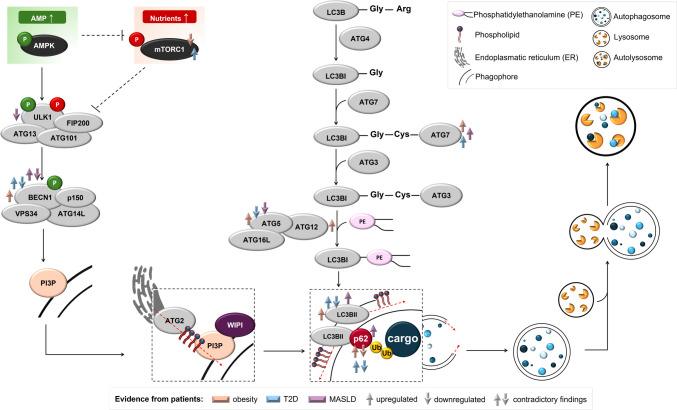
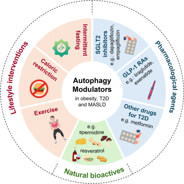
Autophagy is an evolutionarily conserved process that plays a pivotal role in the maintenance of cellular homeostasis and its impairment has been implicated in the pathogenesis of various metabolic diseases including obesity, type 2 diabetes (T2D), and metabolic dysfunction-associated steatotic liver disease (MASLD). This review synthesizes the current evidence from human studies on autophagy alterations under these metabolic conditions. In obesity, most data point to autophagy upregulation during the initiation phase of autophagosome formation, potentially in response to proinflammatory conditions in the adipose tissue. Autophagosome formation appears to be enhanced under hyperglycemic or insulin-resistant conditions in patients with T2D, possibly acting as a compensatory mechanism to eliminate damaged organelles and proteins. Other studies have proposed that prolonged hyperglycemia and disrupted insulin signaling hinder autophagic flux, resulting in the accumulation of dysfunctional cellular components that can contribute to β-cell dysfunction. Evidence from patients with MASLD supports autophagy inhibition in disease progression. Nevertheless, given the available data, it is difficult to ascertain whether autophagy is enhanced or suppressed in these conditions because the levels of autophagy markers depend on the overall metabolism of specific organs, tissues, experimental conditions, or disease duration. Owing to these constraints, determining whether the observed shifts in autophagic activity precede or result from metabolic diseases remains challenging. Additionally, autophagy-modulating strategies are shortly discussed. To conclude, more studies investigating autophagy impairment are required to gain a more comprehensive understanding of its role in the pathogenesis of obesity, T2D, and MASLD and to unveil novel therapeutic strategies for these conditions.
Keywords: Autophagy modulators; Cellular quality control; Metabolic diseases; Patients; Therapies; Tissue biopsy.
© 2024. The Author(s).
Conflict of interest statement
None.
Figures
Similar articles
-
Hepatic steatosis and not type 2 diabetes, body mass index, or hepatic fibrosis associates with hyperglucagonemia in individuals with steatotic liver disease.Am J Physiol Gastrointest Liver Physiol. 2024 Oct 1;327(4):G558-G570. doi: 10.1152/ajpgi.00147.2024. Epub 2024 Aug 6. Am J Physiol Gastrointest Liver Physiol. 2024. PMID: 39104323
-
Autophagy in adipose tissue and the beta cell: implications for obesity and diabetes.Diabetologia. 2014 Aug;57(8):1505-16. doi: 10.1007/s00125-014-3255-3. Epub 2014 May 5. Diabetologia. 2014. PMID: 24795087 Review.
-
Target acquired: Selective autophagy in cardiometabolic disease.Sci Signal. 2017 Feb 28;10(468):eaag2298. doi: 10.1126/scisignal.aag2298. Sci Signal. 2017. PMID: 28246200 Free PMC article. Review.
-
Epidemiology of metabolic dysfunction-associated steatotic liver disease.Clin Mol Hepatol. 2025 Feb;31(Suppl):S32-S50. doi: 10.3350/cmh.2024.0431. Epub 2024 Aug 19. Clin Mol Hepatol. 2025. PMID: 39159948 Free PMC article. Review.
-
The role of adipose tissue dysfunction in hepatic insulin resistance and T2D.J Endocrinol. 2024 Aug 2;262(3):e240115. doi: 10.1530/JOE-24-0115. Print 2024 Sep 1. J Endocrinol. 2024. PMID: 38967989 Free PMC article. Review.
Cited by
-
A Systematic Review of Metabolic Syndrome: Key Correlated Pathologies and Non-Invasive Diagnostic Approaches.J Clin Med. 2024 Oct 2;13(19):5880. doi: 10.3390/jcm13195880. J Clin Med. 2024. PMID: 39407941 Free PMC article. Review.
-
Bacteria in hypertrophic scars promote scar formation through HSBP1-mediated autophagy.Wound Repair Regen. 2025 Jan-Feb;33(1):e13253. doi: 10.1111/wrr.13253. Wound Repair Regen. 2025. PMID: 39823159 Free PMC article.
-
Relationship Between Dietary Nutrient Intake and Autophagy-Related Genes in Obese Humans: A Narrative Review.Nutrients. 2024 Nov 22;16(23):4003. doi: 10.3390/nu16234003. Nutrients. 2024. PMID: 39683397 Free PMC article. Review.
-
Vitamin D3 affects liver expression of pro-/anti-inflammatory cytokines and nitric oxide synthases in type 2 diabetes.Exp Biol Med (Maywood). 2025 Jul 24;250:10456. doi: 10.3389/ebm.2025.10456. eCollection 2025. Exp Biol Med (Maywood). 2025. PMID: 40776976 Free PMC article.
-
Increased autophagy activity suppresses hyperglycemia-related colorectal cancer tumorigenesis both in vitro and in vivo.Am J Cancer Res. 2025 Jul 15;15(7):2949-2969. doi: 10.62347/XSRQ4118. eCollection 2025. Am J Cancer Res. 2025. PMID: 40814362 Free PMC article.
References
-
- Riazi K, Azhari H, Charette JH, Underwood FE, King JA, Afshar EE, Swain MG, Congly SE, Kaplan GG, Shaheen AA (2022) The prevalence and incidence of NAFLD worldwide: a systematic review and meta-analysis. Lancet Gastroenterol Hepatol 7:851–861 - PubMed
-
- Ng M, Fleming T, Robinson M, Thomson B, Graetz N, Margono C, Mullany EC, Biryukov S, Abbafati C, Abera SF, Abraham JP, Abu-Rmeileh NM, Achoki T, AlBuhairan FS, Alemu ZA, Alfonso R, Ali MK, Ali R, Guzman NA, Ammar W, Anwari P, Banerjee A, Barquera S, Basu S, Bennett DA, Bhutta Z, Blore J, Cabral N, Nonato IC, Chang JC, Chowdhury R, Courville KJ, Criqui MH, Cundiff DK, Dabhadkar KC, Dandona L, Davis A, Dayama A, Dharmaratne SD, Ding EL, Durrani AM, Esteghamati A, Farzadfar F, Fay DF, Feigin VL, Flaxman A, Forouzanfar MH, Goto A, Green MA, Gupta R, Hafezi-Nejad N, Hankey GJ, Harewood HC, Havmoeller R, Hay S, Hernandez L, Husseini A, Idrisov BT, Ikeda N, Islami F, Jahangir E, Jassal SK, Jee SH, Jeffreys M, Jonas JB, Kabagambe EK, Khalifa SE, Kengne AP, Khader YS, Khang YH, Kim D, Kimokoti RW, Kinge JM, Kokubo Y, Kosen S, Kwan G, Lai T, Leinsalu M, Li Y, Liang X, Liu S, Logroscino G, Lotufo PALu, Ma Y, Mainoo J, Mensah NK, Merriman GA, Mokdad TR, Moschandreas AH, Naghavi J, Naheed M, Nand A, Narayan D, Nelson KM, Neuhouser EL, Nisar ML, Ohkubo MI, Oti T, Pedroza SO et al (2014) Global, regional, and national prevalence of overweight and obesity in children and adults during 1980–2013: a systematic analysis for the Global Burden of Disease Study 2013. Lancet 384:766–781 - PMC - PubMed
-
- Organization, W. H. (2022) WHO European Regional Obesity Report 2022
Publication types
MeSH terms
Grants and funding
- UMO-2021/43/I/NZ3/00510/Narodowe Centrum Nauki
- 22-04100L/Grantová Agentura České Republiky
- 2017XA5J5N/Ministero dell'Istruzione, dell'Università e della Ricerca
- PRINP202242AFC/Ministero dell'Istruzione, dell'Università e della Ricerca
- PRIN2022ALJN73/Ministero dell'Istruzione, dell'Università e della Ricerca
LinkOut - more resources
Full Text Sources
Medical
