

Deep learning CT reconstruction improves liver metastases detection
- PMID: 38971933
- PMCID: PMC11227486
- DOI: 10.1186/s13244-024-01753-1
Deep learning CT reconstruction improves liver metastases detection
Abstract
Objectives: Detection of liver metastases is crucial for guiding oncological management. Computed tomography through iterative reconstructions is widely used in this indication but has certain limitations. Deep learning image reconstructions (DLIR) use deep neural networks to achieve a significant noise reduction compared to iterative reconstructions. While reports have demonstrated improvements in image quality, their impact on liver metastases detection remains unclear. Our main objective was to determine whether DLIR affects the number of detected liver metastasis. Our secondary objective was to compare metastases conspicuity between the two reconstruction methods.
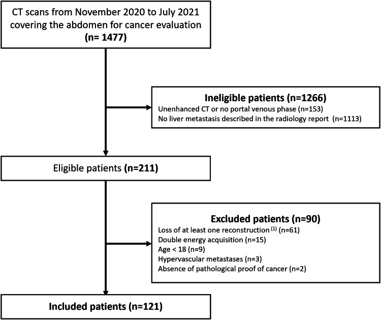
Methods: CT images of 121 patients with liver metastases were reconstructed using a 50% adaptive statistical iterative reconstruction (50%-ASiR-V), and three levels of DLIR (DLIR-low, DLIR-medium, and DLIR-high). For each reconstruction, two double-blinded radiologists counted up to a maximum of ten metastases. Visibility and contour definitions were also assessed. Comparisons between methods for continuous parameters were performed using mixed models.
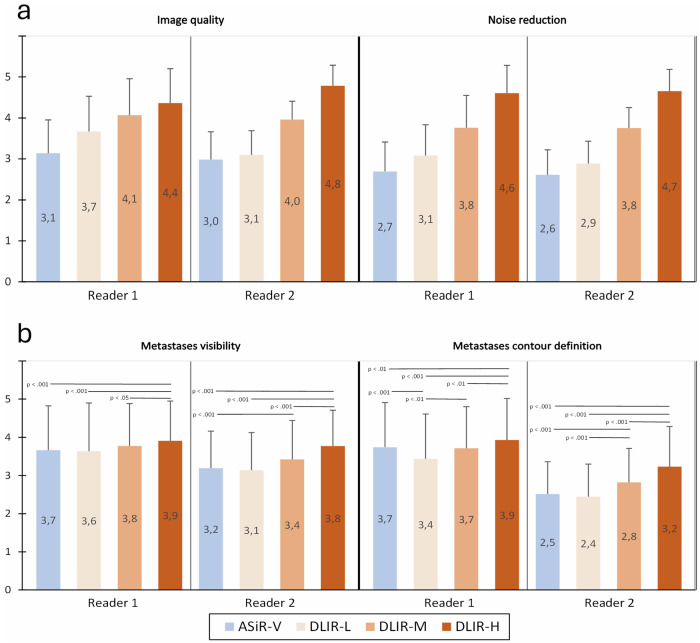
Results: A higher number of metastases was detected by one reader with DLIR-high: 7 (2-10) (median (Q₁-Q₃); total 733) versus 5 (2-10), respectively for DLIR-medium, DLIR-low, and ASiR-V (p < 0.001). Ten patents were detected with more metastases with DLIR-high simultaneously by both readers and a third reader for confirmation. Metastases visibility and contour definition were better with DLIR than ASiR-V.
Conclusion: DLIR-high enhanced the detection and visibility of liver metastases compared to ASiR-V, and also increased the number of liver metastases detected.
Critical relevance statement: Deep learning-based reconstruction at high strength allowed an increase in liver metastases detection compared to hybrid iterative reconstruction and can be used in clinical oncology imaging to help overcome the limitations of CT.
Key points: Detection of liver metastases is crucial but limited with standard CT reconstructions. More liver metastases were detected with deep-learning CT reconstruction compared to iterative reconstruction. Deep learning reconstructions are suitable for hepatic metastases staging and follow-up.
Keywords: Artificial intelligence; Computed tomography; Deep learning; Image reconstruction; Liver neoplasm.
© 2024. The Author(s).
Conflict of interest statement
The authors declare that they have no competing interests.
Figures
References
-
- Germani MM, Borelli B, Boraschi P, et al. The management of colorectal liver metastases amenable of surgical resection: How to shape treatment strategies according to clinical, radiological, pathological and molecular features. Cancer Treat Rev. 2022;106:102382. doi: 10.1016/j.ctrv.2022.102382. - DOI - PubMed
-
- Marion-Audibert A-M, Vullierme M-P, Ronot M, et al. Routine MRI with DWI sequences to detect liver metastases in patients with potentially resectable pancreatic ductal carcinoma and normal liver CT: a prospective multicenter study. AJR. Am J Roentgenol. 2018;211:W217–W225. doi: 10.2214/AJR.18.19640. - DOI - PubMed
LinkOut - more resources
Full Text Sources
