. 2024 Sep:75:103264.
doi: 10.1016/j.redox.2024.103264. Epub 2024 Jul 5.

Redox-dependent plasticity of oxMIF facilitates its interaction with CD74 and therapeutic antibodies

Affiliations

Redox-dependent plasticity of oxMIF facilitates its interaction with CD74 and therapeutic antibodies

Sara Sajko et al. Redox Biol. 2024 Sep.

Abstract

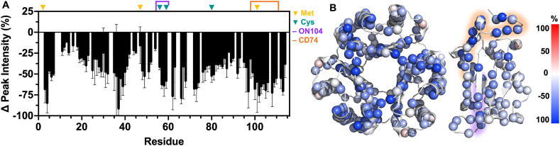
MIF is a ubiquitous protein involved in proinflammatory processes, which undergoes an oxidation-driven conformational change to oxidized (ox)MIF. We demonstrate that hypochlorous acid, produced by neutrophil-released myeloperoxidase (MPO) under inflammatory conditions, effectively oxidizes MIF into the oxMIF isoform, which is specifically recognized by the anti-oxMIF therapeutic antibody, ON104. NMR investigation of MIF oxidized by the MPO system revealed increased flexibility throughout the MIF structure, including at several catalytic and allosteric sites. Mass spectrometry of MPO-oxMIF revealed methionines as the primary site of oxidation, whereas Pro2 and Tyr99/100 remained almost unmodified. ELISA, SPR and cell-based assays demonstrated that structural changes caused by MPO-driven oxidation promoted binding of oxMIF to its receptor, CD74, which does not occur with native MIF. These data reveal the environment and modifications that facilitate interactions between MIF and its pro-inflammatory receptor, and a route for therapeutic intervention targeting the oxMIF isoform.

Keywords: CD74; Hypochlorous acid; Inflammation; MIF; Methionine; Myeloperoxidase; Neutrophils; ON104; oxMIF.


Conflict of interest statement

Declaration of competing interest The authors declare the following financial interests/personal relationships which may be considered as potential competing interests: At the time of the study, SS, MF, AS, IM, JM, GR and MT are employees of OncoOne R&D GmbH (Vienna, Austria), MT, and AS hold shares in OncoOne R&D GmbH, and MT and AS are inventors on a patent application related to the work described here.

Figures

Fig. 1
Fig. 1
MIF and oxMIF are produced in isolated human neutrophils and MPO-oxMIF specifically interacts with ON104. (A, B) Production of MIF and oxMIF by isolated human neutrophils. Neutrophils from 11 healthy donors were stimulated with PMA/ionomycin or left unstimulated (NS; “no stimulation”). 18 h post-stimulation, anti-MIF (A) and anti-oxMIF ELISA (B) were performed on the supernatants. Compared to unstimulated neutrophils, a significant increase in MIF (A) and oxMIF (B) was observed in supernatants from stimulated neutrophils. Data are expressed as mean ± SEM. Statistical analysis was performed using paired and non-parametric Wilcoxon t-test. *p < 0.05. Biological samples n = 11, technical replicates n = 1. (C) Neutrophils from 7 healthy donors, used in (A, B), were also analysed for MPO activity based on colorimetric absorbance at 412 nm (Ellman's assay). Unstimulated neutrophils and those stimulated with PMA/ionomycin are compared. Biological samples n = 7, technical replicates n = 1 per biological sample. Data are reported as mean ± SD. (D) Native MIF, MPO-oxMIF, H2O2-MIF, and NaOCl-oxMIF were tested for ON104 binding by ELISA, which was only observed for MPO-oxMIF (EC50 of 8.1 ± 0.8 nM) and NaOCl-oxMIF (EC50 of 8.8 ± 1.0 nM). Data are normalized by highest MPO-oxMIF binding signal and reported as mean ± SEM of n = 3 independent experiments performed in technical duplicates. (E, F) SPR experiments confirm ON104 binding to MPO-oxMIF, with ON104 used as ligand. (E) Pilot SPR experiments showing MPO-oxMIF binding at 202 nM to immobilized ON104. The absence of binding is apparent for H2O2-MIF at the same concentration, MPO alone, and buffer controls. (F) A KD of 26.6 ± 1.3 nM from a 1:1 Langmuir model was determined from concentration-dependent analyte binding of MPO-oxMIF to immobilized ON104. KD reported as mean ± SD from 3 independent experiments.
Fig. 2
Fig. 2
Intact mass analysis and peptide mass analysis of untreated MIF and MPO-oxMIF. (A) A peak at 12345.98 Da corresponds to the theoretical mass of a MIF monomeric subunit, lacking Met1. Biological samples n = 4, technical replicates n = 1 per biological sample. (B) Treatment of MIF with the MPO-system produced additional peaks with mass increments of +16 Da, due to the addition of one, two or three oxygen atoms. Biological samples n = 5, technical replicates n = 1 per biological sample. (C-E) Peptide mapping MS/MS of MPO-oxMIF where average spectrum m/z values correspond to peptides with oxidized (C) Met3, (D) Met48, and (E) Met102. (CE) Biological samples n = 5, technical replicates n = 1. (AE) One representative spectrum per condition or modification is shown here.
Fig. 3
Fig. 3
NMR analysis of MPO-oxMIF. (A) Changes in NMR 1H15N HSQC resonance intensity (Δ peak intensity) for MPO-oxMIF, calculated relative to untreated MIF. Met and Cys residues are denoted by yellow and teal arrows, respectively. The ON104 binding epitope and purported CD74 binding region are highlighted by purple and orange brackets, respectively. Error bars were determined from n = 3 biological samples, n = 1 technical replicates per sample. (B) The per-residue Δ peak intensity is mapped onto the MIF structure, with blue and red spheres denoting decreases and increases in resonance intensity, respectively. The color gradient represents the magnitude of Δ peak intensity. The ON104 binding epitope and predicted CD74 binding region are highlighted in purple and orange, respectively. (For interpretation of the references to color in this figure legend, the reader is referred to the Web version of this article.)
Fig. 4
Fig. 4
MPO-oxMIF, H2O2-MIF and CD74 binding assays, and proposed impact of HOCl on the MIF trimer. (A) Pilot SPR experiment with 100 nM H2O2-MIF and 100 nM MPO-oxMIF showed that analyte MPO-oxMIF, and not H2O2-MIF, bound to immobilized CD74 ECD. (B) A KD of 11.8 ± 3.7 nM from a 1:1 Langmuir model was extracted from concentration-dependent binding of MPO-oxMIF to CD74 ECD. KD reported as mean ± SD from n = 4 independent experiments. (C) FACS analysis of IFN-γ-treated HT29 cells confirmed the presence of CD74 by anti-CD74-PE antibody. In the absence of IFN-γ, cells did not express CD74. One representative FACS histogram of n = 3 independent experiments is shown. (D) FACS analysis of IFN-γ-treated HT29 cells revealed that the exogenous MPO-oxMIF was detected on the cell surface. MPO-oxMIF was also detected on the surface of non-treated cells. Antibodies used to detect each species are detailed in Materials and Methods. Data are expressed as relative geometric mean fluorescence intensity (MFI) after normalizing to the MFI values of MPO-oxMIF binding to IFN-γ-treated HT-29 detected with anti-MIF mAb and upon the subtraction of the MFI of secondary antibody alone (mean of n = 3 independent experiments ± SEM). (E, F) FACS analysis revealed increased binding of exogenous MPO-oxMIF to CD74-transfected HT29 cells compared to mock-transfected and non-transfected HT29 cells. In contrast, H2O2-MIF does not bind to HT29 cells. Binding was detected with monoclonal anti-MIF antibody (E), or with ON104 (F). Data expressed as relative geometric mean fluorescence intensity (MFI) values after normalizing to the MFI values of MPO-oxMIF binding to CD74-transfected HT-29 detected with anti-MIF mAb and upon the subtraction of the MFI of secondary antibody alone (mean of n = 3 independent experiments ± SEM). (G) Representative FACS histograms of one donor from panel H showing oxMIF presence on the cell surface of unstimulated (NS) versus LPS-activated neutrophils. (H) oxMIF was detected with ON104-AF488 at the cell surface of NS and LPS-activated neutrophils from healthy donors. Data are expressed as a mean ± SEM of n = 5 individual donors. Statistical analysis was performed using paired and non-parametric Wilcoxon t-test. *p < 0.05.

References

    1. Bloom B.R., Bennett B. Mechanism of a reaction in vitro associated with delayed-type hypersensitivity. Science. 1966;153(3731):80–82. doi: 10.1126/science.153.3731.80. - DOI - PubMed
    1. Weiser W.Y., Templet P.A., Witek-Giannottit J.S., Remold H.G., Clarkt S.C., David J.R. Molecular cloning of a cDNA encoding a human macrophage migration inhibitory factor. Immunology. 1989;86 doi: 10.1073/pnas.86.19.7522. - DOI - PMC - PubMed
    1. David J.R. Delayed hypersensitivity in vitro: its mediation by cell-free substances formed by lymphoid cell-antigen interaction. Proc. Natl. Acad. Sci. 1966;56(1):72–77. doi: 10.1073/pnas.56.1.72. - DOI - PMC - PubMed
    1. Calandra T., Roger T. Macrophage migration inhibitory factor: a regulator of innate immunity. Nat. Rev. Immunol. 2003;3(10):791–800. doi: 10.1038/nri1200. - DOI - PMC - PubMed
    1. Kang I., Bucala R. Nature Reviews Rheumatology. Vol. 15. Nature Publishing Group; 2019. The immunobiology of MIF: function, genetics and prospects for precision medicine; pp. 427–437. Issue 7. - DOI - PubMed

MeSH terms

Substances

LinkOut - more resources