Review
. 2024 Aug;40(3):325-336.
doi: 10.1016/j.hcl.2024.04.003.

Advances in Imaging of Compressive Neuropathies

Affiliations
Review

Advances in Imaging of Compressive Neuropathies

Elizabeth A Graesser et al. Hand Clin. 2024 Aug.

Abstract

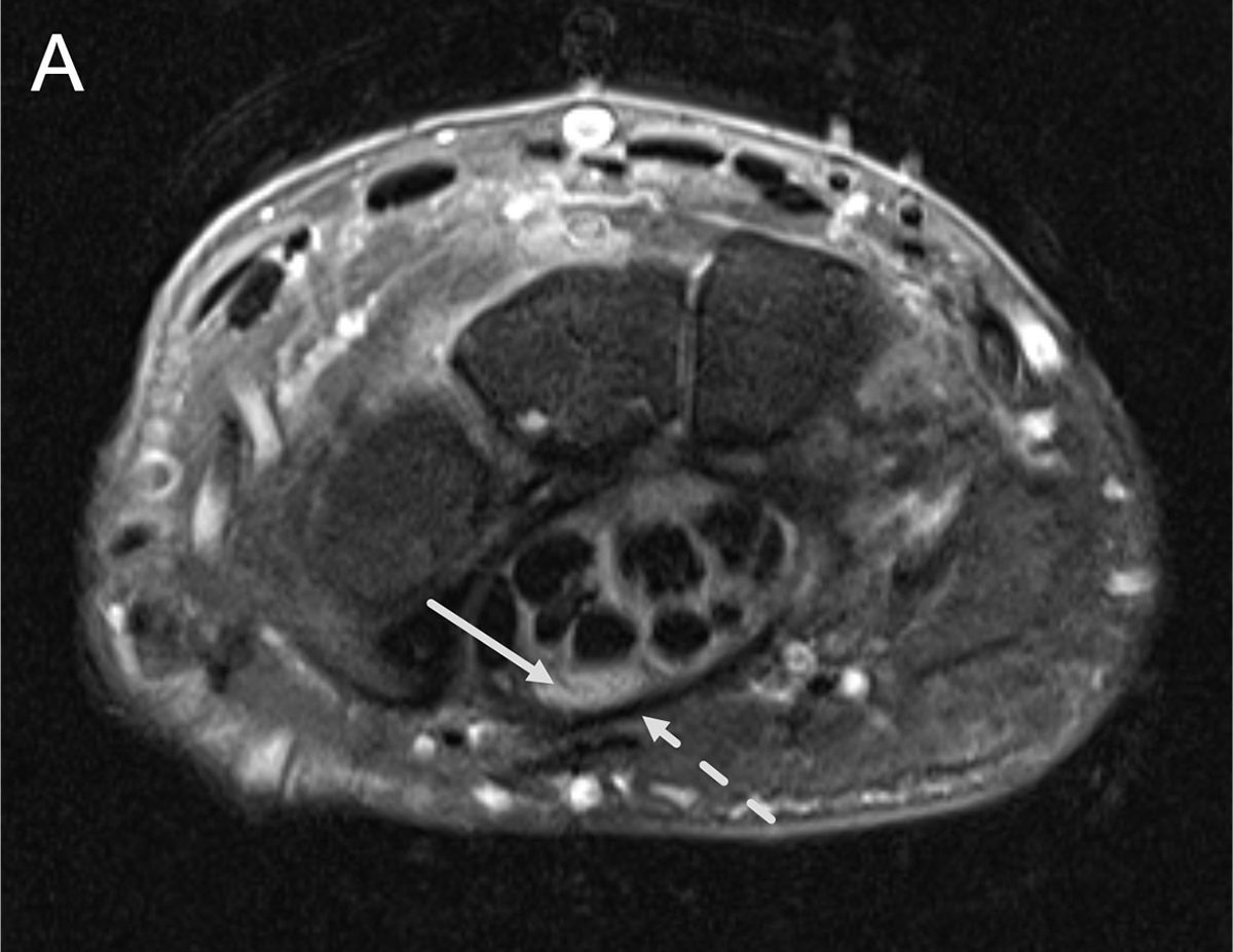
Ultrasound and magnetic resonance neurography are useful modalities to aid in the assessment of compressive neuropathies, although they are still limited in their resolution of nerve microstructure and their capacity to monitor postoperative nerve recovery. Optical coherence tomography, a preclinical imaging modality, is promising in its ability to better identify structural and potential physiologic changes to peripheral nerves, but requires additional testing and research prior to widespread clinical implementation. Further advances in nerve imaging may elucidate the ability to visualize the zone of nerve injury intraoperatively, monitor the progression of nerve regeneration, and localize problems during nerve recovery.

Keywords: Compressive neuropathy; Magnetic resonance neurography; Nerve imaging; Peripheral nerve; Ultrasound.


Conflict of interest statement

Disclosure Dr C.J. Dy is on the Medical Advisory Board for Orthocell. He receives speakers’ fees from Johnson and Johnson/DePuy Synthes. He has research support from the National Institutes of Health and Sonex Healthcare. Dr D.M. Brogan serves as a committee chair in the American Society for Surgery of the Hand and an officer of the Missouri State Orthopedic Association. He receives royalties from Springer and is a paid consultant for Checkpoint Surgical. He has additional research funding from the National Institutes of Health, Department of Defense, United States, American Foundation for Surgery of the Hand, United States, Orthopedic Research and Education Foundation, United States,Checkpoint Surgical, and Neuraptive Therapeutics.

Figures

Figure 1 –
Figure 1 –
Short-axis ultrasound images of a normal median nerve at the carpal tunnel (A) with a cross-sectional area of 9mm2 and a normal ulnar nerve at the elbow (B) with a cross-sectional area of 7mm2. Images courtesy of and copyright protected by Nathan Olafsen MD – used with permission.
Figure 1 –
Figure 1 –
Short-axis ultrasound images of a normal median nerve at the carpal tunnel (A) with a cross-sectional area of 9mm2 and a normal ulnar nerve at the elbow (B) with a cross-sectional area of 7mm2. Images courtesy of and copyright protected by Nathan Olafsen MD – used with permission.
Figure 2 –
Figure 2 –
Ultrasound images demonstrating median neuropathy with a short-axis ultrasound image (A) of an enlarged median nerve in the distal carpal tunnel with a cross-sectional area of 27mm2 and a long-axis ultrasound image (B) of an entrapped median nerve. Images courtesy of and copyright protected by Nathan Olafsen MD – used with permission.
Figure 2 –
Figure 2 –
Ultrasound images demonstrating median neuropathy with a short-axis ultrasound image (A) of an enlarged median nerve in the distal carpal tunnel with a cross-sectional area of 27mm2 and a long-axis ultrasound image (B) of an entrapped median nerve. Images courtesy of and copyright protected by Nathan Olafsen MD – used with permission.
Figure 3 –
Figure 3 –
Short-axis ultrasound image of the ulnar nerve at the elbow demonstrating ulnar neuropathy with an increased cross-sectional area of 14mm2. Image courtesy of and copyright protected by Nathan Olafsen MD – used with permission.
Figure 4 –
Figure 4 –
Coronal 3D STIR (A) and sagittal TIRM (B-E) images of a normal left brachial plexus. The coronal 3D STIR demonstrates normal brachial plexus components extending from the roots to the cords. The sagittal TIRM images illustrates the roots (B), trunks (C), divisions (D), and cords (E) of the brachial plexus. Images courtesy of and copyright protected by Matthew S Parsons MD – used with permission.
Figure 4 –
Figure 4 –
Coronal 3D STIR (A) and sagittal TIRM (B-E) images of a normal left brachial plexus. The coronal 3D STIR demonstrates normal brachial plexus components extending from the roots to the cords. The sagittal TIRM images illustrates the roots (B), trunks (C), divisions (D), and cords (E) of the brachial plexus. Images courtesy of and copyright protected by Matthew S Parsons MD – used with permission.
Figure 5 –
Figure 5 –
Axial T2-weighted fat-saturated (A and B) and sagittal T2-weighted (C) images of the right wrist. The axial images show focal enlargement and T2 hyperintensity of the median nerve (arrows) along with bowing of the flexor retinaculum (dashed arrow). Sagittal T2 (C) weighted images also show focal enlargement and T2 hyperintensity of the median nerve (arrow). Images courtesy of and copyright protected by Matthew S Parsons MD – used with permission.
Figure 5 –
Figure 5 –
Axial T2-weighted fat-saturated (A and B) and sagittal T2-weighted (C) images of the right wrist. The axial images show focal enlargement and T2 hyperintensity of the median nerve (arrows) along with bowing of the flexor retinaculum (dashed arrow). Sagittal T2 (C) weighted images also show focal enlargement and T2 hyperintensity of the median nerve (arrow). Images courtesy of and copyright protected by Matthew S Parsons MD – used with permission.
Figure 6 –
Figure 6 –
Axial T2-weighted fat-saturated (A), axial T1-weighted (B), and oblique coronal (C) and oblique sagittal (D) T2-weighted fat-saturated images of the right elbow. The axial images (A and B) show focal enlargement of the ulnar nerve (arrows) as it courses through the cubital tunnel. The sagittal T2 images (C and D) also show focal enlargement and T2-hyperintensity of the ulnar nerve (arrows). Images courtesy of and copyright protected by Matthew S Parsons MD – used with permission.
Figure 6 –
Figure 6 –
Axial T2-weighted fat-saturated (A), axial T1-weighted (B), and oblique coronal (C) and oblique sagittal (D) T2-weighted fat-saturated images of the right elbow. The axial images (A and B) show focal enlargement of the ulnar nerve (arrows) as it courses through the cubital tunnel. The sagittal T2 images (C and D) also show focal enlargement and T2-hyperintensity of the ulnar nerve (arrows). Images courtesy of and copyright protected by Matthew S Parsons MD – used with permission.

Similar articles

Cited by

References

    1. Demino C, Fowler JR. The Sensitivity and Specificity of Nerve Conduction Studies for Diagnosis of Carpal Tunnel Syndrome: A Systematic Review. Hand. 2021;16(2):174–178. doi: 10.1177/1558944719855442 - DOI - PMC - PubMed
    1. Richardson JK, Evans JE, Warner JH. Information effect on the perception of pain during electromyography. Arch Phys Med Rehabil. 1994;75(6):671–675. - PubMed
    1. London ZN, Burke JF, Hazan R, Hastings MM, Callaghan BC. Electromyography-related pain: Muscle selection is the key modifiable study characteristic. Muscle and Nerve. 2014;49(4):570–574. doi: 10.1002/mus.23974 - DOI - PubMed
    1. Sears ED, Swiatek PR, Hou H, Chung KC. Utilization of Preoperative Electrodiagnostic Studies for Carpal Tunnel Syndrome: An Analysis of National Practice Patterns. J Hand Surg Am. 2016;41(6):665–672.e1. doi: 10.1016/j.jhsa.2016.03.002 - DOI - PMC - PubMed
    1. Dy CJ, Colorado BS, Landau AJ, Brogan DM. Interpretation of Electrodiagnostic Studies: How to Apply It to the Practice of Orthopaedic Surgery. J Am Acad Orthop Surg. 2021;29(13):e646–e654. doi: 10.5435/JAAOS-D-20-00322 - DOI - PMC - PubMed

MeSH terms