. 2024 Dec;13(1):2377599.
doi: 10.1080/22221751.2024.2377599. Epub 2024 Jul 18.

The antibodies against the A137R protein drive antibody-dependent enhancement of African swine fever virus infection in porcine alveolar macrophages

Affiliations

The antibodies against the A137R protein drive antibody-dependent enhancement of African swine fever virus infection in porcine alveolar macrophages

Xiaoke Yang et al. Emerg Microbes Infect. 2024 Dec.

Abstract

African swine fever virus (ASFV) is the causative agent of African swine fever (ASF), a highly contagious disease that can kill up to 100% of domestic pigs and wild boars. It has been shown that the pigs inoculated with some ASF vaccine candidates display more severe clinical signs and die earlier than do pigs not immunized. We hypothesize that antibody-dependent enhancement (ADE) of ASFV infection may be caused by the presence of some unidentified antibodies. In this study, we found that the ASFV-encoded structural protein A137R (pA137R) can be recognized by the anti-ASFV positive sera, indicating that the anti-pA137R antibodies are induced in the ASFV-infected pigs. Interestingly, our results demonstrated that the anti-pA137R antibodies produced in rabbits or pigs enhanced viral replication of different ASFV strains in primary porcine alveolar macrophages (PAMs), the target cells of ASFV. Mechanistic investigations revealed that anti-pA137R antibodies were able to promote the attachment of ASFV to PAMs and two types of Fc gamma receptors (FcγRs), FcγRII and FcγRIII, mediated the ADE of ASFV infection. Taken together, anti-pA137R antibodies are able to drive ASFV ADE in PAMs. These findings shed new light on the roles of anti-ASFV antibodies and have implications for the pathophysiology of the disease and the development of ASF vaccines.

Keywords: A137R protein; African swine fever virus; Fc gamma receptors; antibody-dependent enhancement; viral replication.


Conflict of interest statement

No potential conflict of interest was reported by the author(s).

Figures

Figure 1.
Figure 1.
Anti-pA137R antibodies have the potential to enhance viral infection. (A) Identification of the new ASFV immunogenic proteins. The reactivity of ASFV structural proteins with the convalescent sera was analyzed by western blotting. (B) Mouse anti-pA137R sera augment ASFV infection. ASFV-ΔCD2v-EGFP was incubated with antibodies against the novel immunogenic proteins or the negative sera for 90 min. At 48 h postinfection, the fluorescent cells infected with ASFV-ΔCD2v-EGFP were observed under a fluorescent microscope.
Figure 2.
Figure 2.
Anti-pA137R antibodies are detectable in the convalescent sera from ASFV-infected pigs. (A) The ASFV pA137R specifically reacts with the convalescent sera from the pigs that survived ASFV infection, while the non-convalescent sera from the ASFV-infected pigs were used as a negative control. HEK293 T cells were transfected with pFlag-A137R (2 μg), pFlag-p30 (2 μg), or pCAGGS-Flag (serving as a negative control) for 48 h and then the fluorescence was analyzed by IFA. (B) The reactivity of the purified pA137R with ASFV-convalescent or non-convalescent sera examined by western blotting. The p30 and Cap proteins were used as positive and negative controls, respectively. (C) Anti-pA137R antibodies are induced in ASFV-infected convalescent pigs. The antibodies against pA137R were tested in the sera collected from the ASFV-infected pigs by the indirect ELISA using the recombinant pA137R or GST as coated antigens. (D) Subviral localization of pA137R. ASFV-infected PAMs were fixed at 18 h postinfection and immunolabelled with a rabbit anti-pA137R antibodies followed by an anti-rabbit antibody conjugated to 5-nm-diameter gold particles. The arrowheads indicate the gold particles present on outer protein capsid of intracellular virus particles and the capsid is depicted in black line. Bar, 200 nm. (E) Multiple sequence alignment of pA137R of diverse ASFV isolates. A conservation analysis of the pA137R among genotypes I, II, and I/II recombinant ASFV isolates was conducted using the Jalview and ClustalW algorithms.
Figure 3.
Figure 3.
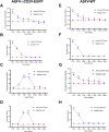
Rabbit anti-pA137R antibodies induce the ASFV ADE in PAMs. (A and B) Increased ASFV-ΔCD2v-EGFP replication by rabbit anti-pA137R antibodies of dilutions from 1:50 to 1:3200 in PAMs. PAMs were infected with ASFV-ΔCD2v-EGFP in the presence or absence of anti-pA137R antibodies. The viral genome copies were quantified by qPCR, and the viral titers (Log10 TCID50/mL) were titrated at 48 h postinfection (hpi). (C and D) Enhanced ASFV-ΔCD2v-EGFP replication by anti-pA137R antibodies at dilutions from 1:10 to 1:640 in PAMs. The viral genome copies, and viral titers (Log10 TCID50/mL) were determined following infection with the complex of ASFV-ΔCD2v-EGFP and the sera. (E and F) Enhanced ASFV-WT replication by anti-pA137R antibodies in PAMs. The replication of ASFV-WT in the presence of anti-pA137R antibodies with serial dilutions from 1:50 to 1:3200 was examined. The viral genome copies and viral titers (Log10 HAD50/mL) of ASFV-WT were determined at 48 hpi. (G and H) Anti-pA137R antibodies at high concentration enhanced ASFV-WT infection in PAMs. The experimental data of each group were separately recorded under the treatment of a higher concentration of anti-pA137R antibodies with serial dilutions from 1:10 to 1:640. The viral genome copies were quantified by qPCR, and the viral titers of ASFV-WT were titrated as Log10 HAD50/mL at 48 hpi. Specific-pathogen-free rabbit sera were used as a negative control. The data represented three independent experiments. All the data were expressed as fold changes relative to the control. The error bars represented the standard errors of the means. All the data were analyzed using the Student’s t test: ***, P < 0.001; **, P < 0.01; *, P < 0.05; ns, not significant, P > 0.05.
Figure 4.
Figure 4.
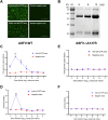
Porcine anti-pA137R antibodies drive ADE of ASFV-WT but not of ASFV-ΔA137R in PAMs. (A and B) The reactivity and purity of the purified anti-pA137R antibodies. (C and D) Porcine anti-pA137R antibodies increase ASFV-WT replication in PAMs. Anti-pA137R antibodies and negative sera at 10-fold serial dilutions starting 1:10 were incubated with ASFV-WT, and the mix were added to PAMs to determine the enhancement folds of the viral genome copies and titers. (E and F) Anti-pA137R antibodies do not affect the replication of ASFV-ΔA137R in PAMs. After a 1.5-h incubation of anti-pA137R antibodies or negative sera of serial dilutions with ASFV-ΔA137R, the viral genome copies and titers of each group were determined. Specific-pathogen-free porcine sera were used as a negative control. The data represented three independent experiments. The error bars represent the standard errors of the means. All the data were analyzed using the Student’s t test: ***, P < 0.001; **, P < 0.01; *, P < 0.05; ns, not significant, P > 0.05.
Figure 5.
Figure 5.
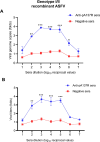
Porcine anti-pA137R antibodies drive ADE of the genotype I/II recombinant ASFV strain. (A and B) The genotype I/II recombinant ASFV strain was incubated with anti-pA137R antibodies of different concentrations. Specific-pathogen-free pig sera were used as a negative control. The mixture was added to PAMs to determine the enhancement folds of the viral genome copies and titers. The data represented three independent experiments. The error bars represent the standard errors of the means. All the data were analyzed using the Student’s t test: ***, P < 0.001; **, P < 0.01; *, P < 0.05; ns, no significance.
Figure 6.
Figure 6.
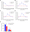
Porcine anti-pA137R antibodies promote the attachment of ASFV-WT to PAMs. (AD) Rabbit anti-pA137R antibodies facilitate the attachment of ASFV-WT to PAMs. To check the effects of anti-pA137R antibodies of serial dilutions on the attachment of ASFV to the target cells, PAMs were infected with ASFV-WT at an MOI of 1, 3, or 5 in the presence or absence of anti-pA137R antibodies of serial dilutions. After a 2-h incubation at 4°C, the ASFV genomic DNA was extracted and quantified by qPCR (the results were shown as fold changes). (E) Porcine anti-pA137R antibodies promoted the attachment of ASFV-WT but not ASFV-ΔA137R to PAMs. The viral genome copies of ASFV-WT and ASFV-ΔA137R in the presence of porcine anti-pA137R antibodies or negative sera of 1:100 dilution were quantified by qPCR. The data represented three independent experiments. The error bars represented the standard errors of the means. All the data were analyzed using the Student’s t test: ***, P < 0.001; **, P < 0.01; *, P < 0.05; ns, not significant, P > 0.05.
Figure 7.
Figure 7.
FcγRII and FcγRIII are involved in the ASFV ADE. (A–C) Inhibition of ASFV ADE in the presence of anti-CD16/CD32 antibodies in a dose-dependent manner. Using different amounts of anti-CD16/CD32 antibodies to block the FcγRII and FcγRIII on the surface of PAMs, subsequently the PAMs were infected using the complex of ASFV-WT and anti-pA137R antibodies and incubated for 24 h. (D–I) ADE of ASFV infection is mainly mediated by FcγRII or FcγRIII. Different amounts of anti-CD16 or anti-CD32 antibodies were inoculated with PAMs. ASFV-WT in the presence of porcine anti-pA137R antibodies or negative sera of 1:100 dilution was added to the PAMs. At 24 h postinfection, the viral genome copies were quantified by qPCR, and the viral titers of ASFV-WT were titrated as Log10 HAD50/mL. The data represented three independent experiments. The error bars represented the standard errors of the means. All the data were analyzed using the Student’s t test: ***, P < 0.001; **, P < 0.01; *, P < 0.05; ns, not significant, P > 0.05.

Similar articles

Cited by

References

    1. Bailey MJ, Broecker F, Freyn AW, et al. . Human monoclonal antibodies potently neutralize Zika virus and select for escape mutations on the lateral ridge of the envelope protein. J Virol. 2019;93:e00405–19. doi:10.1128/JVI.00405-19 - DOI - PMC - PubMed
    1. Vanderven HA, Jegaskanda S, Wheatley AK, et al. . Antibody-dependent cellular cytotoxicity and influenza virus. Curr Opin Virol. 2017;22:89–96. doi:10.1016/j.coviro.2016.12.002 - DOI - PubMed
    1. Ye ZW, Yuan S, Poon KM, et al. . Antibody-dependent cell-mediated cytotoxicity epitopes on the hemagglutinin head region of pandemic H1N1 influenza virus play detrimental roles in H1N1-infected mice. Front Immunol. 2017;8:317. - PMC - PubMed
    1. Hawkes RA. Enhancement of the infectivity of Arboviruses by specific antisera produced in domestic fowls. Aust J Exp Biol Med Sci. 1964;42:465–482. doi:10.1038/icb.1964.44 - DOI - PubMed
    1. Sicca F, Neppelenbroek S, Huckriede A.. Effector mechanisms of influenza specific antibodies: neutralization and beyond. Expert Rev Vaccines. 2018;17:785–795. doi:10.1080/14760584.2018.1516553 - DOI - PubMed

LinkOut - more resources