. 2024 May 28;9(13):e178216.
doi: 10.1172/jci.insight.178216.

IL-10 inhibition during immunization improves vaccine-induced protection against Staphylococcus aureus infection

Affiliations

IL-10 inhibition during immunization improves vaccine-induced protection against Staphylococcus aureus infection

Alanna M Kelly et al. JCI Insight. .

Abstract

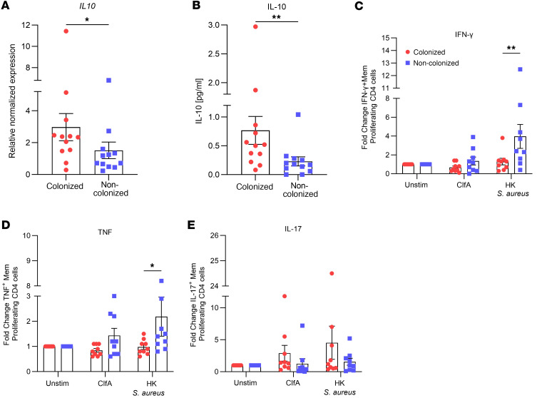
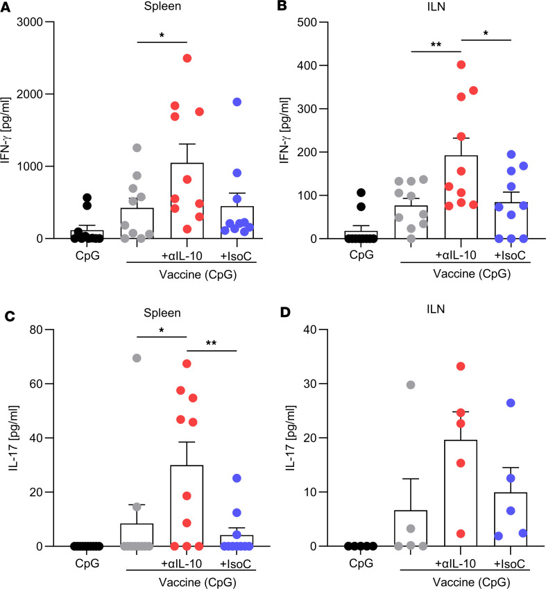
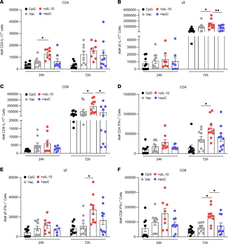
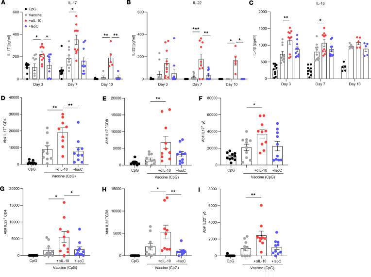
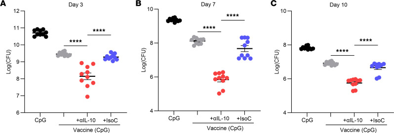
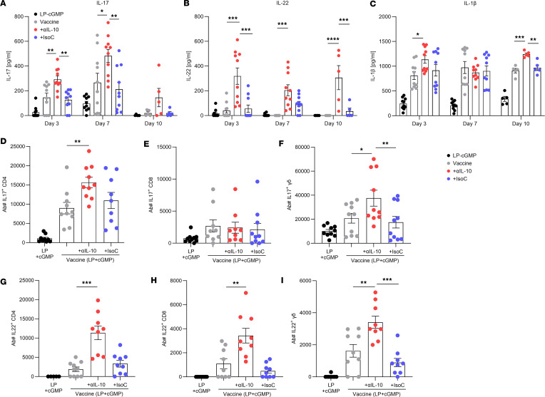
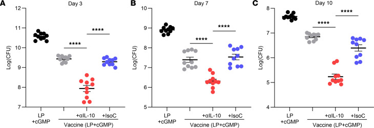
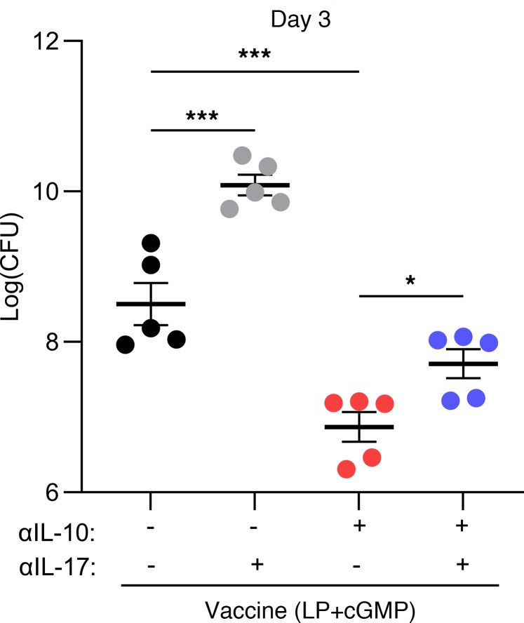
Staphylococcus aureus is a major human pathogen. An effective anti-S. aureus vaccine remains elusive as the correlates of protection are ill-defined. Targeting specific T cell populations is an important strategy for improving anti-S. aureus vaccine efficacy. Potential bottlenecks that remain are S. aureus-induced immunosuppression and the impact this might have on vaccine-induced immunity. S. aureus induces IL-10, which impedes effector T cell responses, facilitating persistence during both colonization and infection. Thus, it was hypothesized that transient targeting of IL-10 might represent an innovative way to improve vaccine efficacy. In this study, IL-10 expression was elevated in the nares of persistent carriers of S. aureus, and this was associated with reduced systemic S. aureus-specific Th1 responses. This suggests that systemic responses are remodeled because of commensal exposure to S. aureus, which negatively implicates vaccine function. To provide proof of concept that targeting immunosuppressive responses during immunization may be a useful approach to improve vaccine efficacy, we immunized mice with T cell-activating vaccines in combination with IL-10-neutralizing antibodies. Blocking IL-10 during vaccination enhanced effector T cell responses and improved bacterial clearance during subsequent systemic and subcutaneous infection. Taken together, these results reveal a potentially novel strategy for improving anti-S. aureus vaccine efficacy.

Keywords: Bacterial infections; Bacterial vaccines; Immunology; T cells; Vaccines.


Conflict of interest statement

Conflict of interest: KHGM is an inventor on a patent (US 2023285531A1) concerning LP1569 as a vaccine adjuvant.

Figures

Figure 1
Figure 1. Persistent nasal colonization with S. aureus is associated with enhanced IL-10 responses locally within the nasal tissue and altered systemic T cell responses upon S. aureus reexposure.
Persistently colonized individuals were identified as those who had 3 consecutive nasal swab cultures positive for S. aureus over a 6-week period. Individuals who tested negative for each swab culture were classified as “noncolonized.” Nasal mucosa was swabbed, and RNA was extracted. IL10 gene expression levels were assessed using quantitative reverse transcription PCR (A). The mRNA values were expressed as mean relative expression ± SEM and compared with baseline IL-10 expression from noncolonized individuals after normalizing to β-actin RNA expression. (Experimental unit = 1 donor, n = 12/group.) Mucosal lining fluid (MLF) was collected using Nasosorption FX·i devices, and IL-10 concentration was measured using a V-plex multiplex ELISA (B). Results are expressed as mean protein expression ± SEM. (Experimental unit = 1 donor, n = 12/group.) Purified CD4+ T cells were carboxyfluorescein diacetate succinimidyl ester–labeled (CFSE-labeled) and cocultured with autologous irradiated antigen-presenting cells from a subgroup of persistently colonized or noncolonized individuals. Cells were stimulated with media alone, ClfA (0.88 μM), or heat-killed S. aureus Newman strain (1 μg/mL) for 8 days. The proportions of IFN-γ+ (C), TNF+ (D), and IL-17+ (E) proliferating memory CD45RO+CD4+ T cells were then assessed. Values are expressed as mean fold-change ± SEM. (Experimental unit = 1 donor, n = 9/group.) Statistical analysis was carried out by Mann-Whitney U test to analyze variances between groups for gene expression and ELISA data or 2-way ANOVA with Holm-Šídák posttest for flow cytometry data. *P ≤ 0.05; **P ≤ 0.01.
Figure 2
Figure 2. Immunization in the presence of anti–IL-10 increases ClfA-specific IFN-γ and IL-17 production by T cells of the spleen and ILNs.
Mice were immunized with CpG (50 μg); vaccine only, consisting of CpG + ClfA (5 μg); vaccine + anti–IL-10 (150 μg); or vaccine + isotype control (150 μg) via s.c. injection on day 0, 14, and 28. On day 42 ILNs and spleen were removed, and ClfA-specific responses were assessed by ex vivo stimulation with media only or ClfA (5 μg/mL) for 72 hours. The levels of IFN-γ in the spleen (A) and ILNs (B), and IL-17 in the spleen (C) and ILNs (D), were determined by ELISA. ClfA-specific responses were determined by subtracting responses to media alone. Results are expressed as mean ± SEM. (Experimental unit = 1 mouse, n =5–10/group, total mice used 40, experiment was performed twice.) Statistical analysis was carried out by 1-way ANOVA with Tukey posttest. *P ≤ 0.05, **P ≤ 0.01.
Figure 3
Figure 3. IL-10 inhibition during vaccination with a CpG-based S. aureus vaccine improves T cell immune responses during systemic infection.
Mice were immunized with CpG (50 μg); vaccine only, consisting of CpG + ClfA (5 μg); vaccine + anti–IL-10 (150 μg); or vaccine + isotype control (150 μg) via s.c. injection on day 0, 14, and 28. On day 42 mice were challenged with S. aureus strain PS80 (5 × 10⁸ CFU) via i.p. injection. At 24 hours and 72 hours after infection, cells of the peritoneal cavity were isolated to assess the number of IL-17+ CD4+ (A), γδ (B), and CD8+ (C) T cells and the number of IFN-γ+ CD4+ (D), γδ (E), and CD8+ (F) T cells in the peritoneum by flow cytometry. Results are expressed as mean absolute number of cell type ± SEM. (Experimental unit = 1 mouse, n = 7–10/group total mice used; 80, experiment was performed twice.) Statistical analysis was carried out by 2-way ANOVA with Holm-Šídák posttest. *P ≤ 0.05, **P ≤ 0.01.
Figure 4
Figure 4. IL-10 inhibition during vaccination with a CpG-based S. aureus vaccine improves bacterial clearance during systemic infection.
Mice were immunized with CpG (50 μg); vaccine only, consisting of CpG + ClfA (5 μg); vaccine + anti–IL-10 (150 μg); or vaccine + isotype control (150 μg) via s.c. injection on day 0, 14, and 28. On day 42 mice were challenged with S. aureus strain PS80 (5 × 10⁸ CFU) via i.p. injection. At 24 hours and 72 hours after infection bacterial burden was assessed in the peritoneal cavity (A), blood (B), kidney (C), and spleen (D). Results are expressed as Log(CFU) ± SEM. (Experimental unit = 1 mouse, n = 5/group, total mice used 40, experiment was performed once.) Statistical analysis was carried out by 2-way ANOVA with Holm-Šídák posttest. *P ≤ 0.05, **P ≤ 0.01, ***P ≤ 0.001, ****P ≤ 0.0001.
Figure 5
Figure 5. IL-10 inhibition during vaccination improves T cell immune responses during a subsequent S. aureus s.c. infection.
Mice were immunized with CpG (50 μg); vaccine only, consisting of CpG + ClfA (5 μg); vaccine + anti–IL-10 (150 μg); or vaccine + isotype control (150 μg). All injections were via s.c. injection on day 0, 14, and 28. On day 42 mice were s.c. infected with S. aureus USA300 (LAC) (2 × 10⁷ CFU). On day 3, 7, and 10 after infection, an 8 mm skin punch biopsy was taken at the infection site and homogenized, and undiluted homogenate supernatants were used for IL-17 (A), IL-22 (B), and IL-1β (C) cytokine production analysis by ELISA. Results are expressed as mean protein expression ± SEM. (Experimental unit = 1 mouse, n = 5–10/group, total mice used 100, experiment was performed twice.) At 72 hours after infection cells of the abscess were isolated to assess the number of IL-17+ CD4+ (D), CD8+ (E), and γδ (F) T cells and the number of IL-22+ CD4+ (G), CD8+ (H), and γδ (I) T cell subtypes by flow cytometry. Results are expressed as mean absolute number of cell type ± SEM. (Experimental unit = 1 mouse, n = 9–10/group, total mice used 40, experiment was performed twice.) Statistical analysis was carried out by 1-way ANOVA with Tukey posttest for flow cytometry data or 2-way ANOVA with Holm-Šídák posttest for ELISA data. *P ≤ 0.05; **P ≤ 0.01, ***P ≤ 0.001.
Figure 6
Figure 6. IL-10 inhibition during vaccination improves clearance of S. aureus during a subsequent s.c. infection.
Mice were immunized with CpG (50 μg); vaccine only, consisting of CpG + ClfA (5 μg); vaccine + anti–IL-10 (150 μg); or vaccine + isotype control (150 μg). All injections were via s.c. injection on day 0, 14, and 28. On day 42 mice were s.c. infected with S. aureus USA300 (LAC) (2 × 10⁷ CFU). On day 3 (A), 7 (B), and 10 (C) after infection, an 8 mm skin punch biopsy was taken at the infection site and homogenized and bacterial burden was assessed. Results are expressed as Log(CFU) ± SEM. (Experimental unit = 1 mouse, n = 10/group, total mice used 120, experiment was performed twice.) Statistical analysis was carried out by 1-way ANOVA with Tukey posttest. ****P ≤ 0.0001.
Figure 7
Figure 7. IL-10 inhibition during vaccination with a novel adjuvant improves T cell immune responses against a s.c. S. aureus infection.
Mice were immunized with LP1569 (50 μg) + cGMP (10 μg); vaccine only, consisting of LP1569 + cGMP + ClfA (5 μg); vaccine + anti–IL-10 (150 μg); or vaccine + isotype control (150 μg). All injections were via s.c. injection on day 0, 14, and 28. On day 42 mice were s.c. infected with S. aureus USA300 (LAC) (2 × 10⁷ CFU). On day 3, 7, and 10 after infection, an 8 mm skin punch biopsy was taken at the infection site and homogenized, and undiluted homogenate supernatants were then used for IL-17 (A), IL-22 (B), and IL-1β (C) cytokine production analysis by ELISA. Results are expressed as mean protein expression ± SEM. (Experimental unit = 1 mouse, n = 5–10/group, total mice used 100, experiment was performed twice.) At 72 hours postinfection cells were isolated from the skin to assess the number of IL-17+ CD4+ (D), CD8+ (E), and γδ (F) T cells and the number of IL-22+ CD4+ (G), CD8+ (H), and γδ (I) T cells by flow cytometry. Results are expressed as mean absolute number of cell type ± SEM. (Experimental unit = 1 mouse, n = 9–10/group, total mice used 40, experiment was performed twice.) Statistical analysis was carried out by 1-way ANOVA with Tukey posttest for flow cytometry data or 2-way ANOVA with Holm-Šídák posttest. *P ≤ 0.05, **P ≤ 0.01, ***P ≤ 0.001, ****P ≤ 0.0001.
Figure 8
Figure 8. IL-10 inhibition during vaccination with a novel adjuvant improves clearance of S. aureus during a subsequent s.c. infection.
Mice were immunized with LP1569 (50 μg) + cGMP (10 μg); vaccine only, consisting of LP1569 + cGMP + ClfA (5 μg); vaccine + anti–IL-10 (150 μg); or vaccine + isotype control (150 μg). All injections were via s.c. injection on day 0, 14, and 28. On day 42 mice were s.c. infected with S. aureus USA300 (LAC) (2 × 10⁷ CFU). On day 3 (A), 7 (B), and 10 (C) after infection, an 8 mm skin punch biopsy was taken at the infection site and homogenized and bacterial burden was assessed. Results are expressed as Log(CFU) ± SEM. (Experimental unit = 1 mouse, n = 10/group, total mice used 120). Statistical analysis was carried out by 1-way ANOVA with Tukey posttest. ****P ≤ 0.0001.
Figure 9
Figure 9. IL-17 blocking during s.c. S. aureus infection impedes bacterial clearance and reduces the protective effect of IL-10 inhibition during vaccination.
Mice were immunized with LP1569 (50 μg) + cGMP (10 μg); vaccine only, consisting of LP1569 + cGMP + ClfA (5 μg); vaccine + anti–IL-10 (150 μg); or vaccine + isotype control (150 μg). All injections were via s.c. injection on day 0, 14, and 28. On day 42 mice were s.c. administered anti–IL-17 (50 μg) or isotype control (50 μg) alongside S. aureus USA300 (LAC) (2 × 10⁷ CFU) and again at 24 hours after infection. On day 3 after infection an 8 mm skin punch biopsy was taken at the infection site and homogenized and bacterial burden was assessed. Results are expressed as Log(CFU) ± SEM. (Experimental unit = 1 mouse, n = 5/group, total mice used = 20.) Statistical analysis was carried out by 1-way ANOVA with Tukey posttest. *P ≤ 0.05, ***P ≤ 0.001.

Similar articles

Cited by

References

    1. GBD 2019 Antimicrobial Resistance Collaborators Global mortality associated with 33 bacterial pathogens in 2019: a systematic analysis for the Global Burden of Disease Study 2019. Lancet. 2022;400(10369):2221–2248. doi: 10.1016/S0140-6736(22)02185-7. - DOI - PMC - PubMed
    1. Antimicrobial Resistance C. Global burden of bacterial antimicrobial resistance in 2019: a systematic analysis. Lancet. 2022;399(10325):629–655. doi: 10.1016/S0140-6736(21)02724-0. - DOI - PMC - PubMed
    1. Melzer M, Welch C. Thirty-day mortality in UK patients with community-onset and hospital-acquired meticillin-susceptible Staphylococcus aureus bacteraemia. J Hosp Infect. 2013;84(2):143–150. doi: 10.1016/j.jhin.2012.12.013. - DOI - PubMed
    1. Olaniyi R, et al. Staphylococcus aureus-associated skin and soft tissue infections: anatomical localization, epidemiology, therapy and potential prophylaxis. Curr Top Microbiol Immunol. 2017;409:199–227. doi: 10.1007/82_2016_32. - DOI - PubMed
    1. European Antimicrobial Resistance Collaborators. The burden of bacterial antimicrobial resistance in the WHO European region in 2019: a cross-country systematic analysis. Lancet Public Health. 2022;7(11):e897–e913. doi: 10.1016/S2468-2667(22)00225-0. - DOI - PMC - PubMed