. 2024 Aug;36(35):e2313791.
doi: 10.1002/adma.202313791. Epub 2024 Jul 8.

Expanding RNAi to Kidneys, Lungs, and Spleen via Selective ORgan Targeting (SORT) siRNA Lipid Nanoparticles

Affiliations

Expanding RNAi to Kidneys, Lungs, and Spleen via Selective ORgan Targeting (SORT) siRNA Lipid Nanoparticles

Amogh Vaidya et al. Adv Mater. 2024 Aug.

Abstract

Inhibition of disease-causing mutations using RNA interference (RNAi) has resulted in clinically approved medicines with additional candidates in late stage trials. However, targetable tissues currently in preclinical development are limited to liver following systemic intravenous (IV) administration because predictable delivery of siRNA to non-liver tissues remains an unsolved challenge. Here, evidence of durable extrahepatic gene silencing enabled by siRNA Selective ORgan Targeting lipid nanoparticles (siRNA SORT LNPs) to the kidneys, lungs, and spleen is provided. LNPs excel at dose-dependent silencing of tissue-enriched endogenous targets resulting in 60%-80% maximal knockdown after a single IV injection and up to 88% downregulation of protein expression in mouse lungs after two doses. To examine knockdown potency and unbiased organ targeting, B6.129TdTom/EGFP mice that constitutively express the TdTomato transgene across all cell types are utilized to demonstrate 58%, 45%, and 15% reduction in TdTomato fluorescence in lungs, spleen, and kidneys, respectively. Finally, physiological relevance of siRNA SORT LNP-mediated gene silencing is established via acute suppression of endogenous Tie2 which induces lung-specific phenotypic alteration of vascular endothelial barrier. Due to plethora of extrahepatic diseases that may benefit from RNAi interventions, it is anticipated that the findings will expand preclinical landscape of therapeutic targets beyond the liver.

Keywords: Selective ORgan Targeting (SORT); extrahepatic siRNA silencing; kidney siRNA delivery; lipid nanoparticles (LNPs); unbiased knockdown.


Figures

Figure 1.
Figure 1.
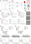
siRNA SORT LNPs possessed amenable physicochemical attributes for delivering active and functional siRNA in vitro and in vivo to extrahepatic tissues. A) Formulation scheme for siRNA SORT LNPs consisting of five lipids. LNPs containing unique SORT lipids delivered functional siRNA to lungs and kidneys (DOTAP-50), liver (DODAP-20), and spleen (18PA-15) in mice. B) Hydrodynamic diameter and polydispersity index (PDI) and C) Zeta potential (mV) for siRNA SORT LNPs. D) RiboGreen assay for estimating % siRNA encapsulation efficiency. E) Representative Cryo-EM microscopy images of siRNA SORT LNPs (top to bottom) unveiled a spherical morphology. F) Representative TNS assay curves for determining the global apparent pKa. A pH value corresponding to 50% TNS fluorescence was attested as the pKa value for the respective LNP formulation. Non-linear best fit line was drawn to estimate the pKa values. G-J) In vitro knockdown efficacy was evaluated 24 h after siRNA SORT LNPs treatment in HeLa-Luc cells with GAPDH and constitutively expressed Luciferase as targets. Cells were treated with SORT LNPs containing 10 nM of either scrambled siRNA (siScrambled), GAPDH siRNA (siGAPDH), or Luciferase siRNA (siLuc). Relative GAPDH activity (G) and Luciferase knockdown efficiency (H) was quantified after 24 h (One-way ANOVA with Tukey’s post hoc test **** p < 0.0001). I) Dose-dependent silencing was performed to understand the dynamic working range of the formulations and analyzed by western blot against GAPDH. The median effective dose (ED50) for all formulations was in the nanomolar range (α-Actinin - internal loading control). J) Dose dependence and cell viability for SORT LNPs containing siLuc was quantified in HeLa-Luc cells using ONE-Glo assay (left and right axis respectively). A sigmoidal silencing curve spanning across three logarithmic scales was observed with ED50 values in the sub-nanomolar range (Non-linear regression curve fit). In general, siRNA SORT LNPs exceled in their silencing capability across both genes tested. Data are shown as mean ± SEM (n=3).
Figure 2.
Figure 2.
siRNA SORT LNPs demonstrated dose-dependent knockdown of tissue-enriched endogenous targets in mouse lungs, kidneys, liver, and spleen at both mRNA and protein levels. A) siRNAs against Tie2, FVII, and CD31 were loaded in DOTAP-50 (Red), DODAP-20 (Blue), and 18PA-15 (Green) LNPs (depicted by pie charts with % molar ratios) respectively to target specific organs; Tie2 (lungs and kidneys), FVII (liver), and CD31 (spleen). Mice were injected with a single dose of DOTAP-50 or 18PA-15 LNPs (0.1, 0.25, 0.5, 1, 1.5 mg kg−1) or DODAP-20 LNPs (0.01, 0.05, 0.1, 0.5, 1 mg kg−1) and respective organs were harvested after 3 days (n=4 mice per dosing group). B-E) mRNA-level knockdown of Tie2, FVII, and CD31 was quantified by qPCR analysis and relative fold change in mRNA was normalized to naïve mice. GAPDH was used as the house-keeping control. F, G, I) Protein-level change after siRNA knockdown of Tie2 and CD31 was measured using western blot. The band intensity was quantified with Image J and a graph of relative expression for every dose was plotted. H) FVII silencing activity of DODAP-20 LNPs was evaluated using a FVII chromogenic kit. EC50 values in B-I were calculated with non-linear regression curve fit analysis. J-L) Representative western blots of Tie2 and CD31 following treatments with DOTAP-50 (lungs, kidneys) and 18PA-15 (spleen). Gene targets are color coded and matched to respective ‘SORT’ lipids in the formulation pie-chart. Data are shown as mean ± SEM (n=4).
Figure 3.
Figure 3.
siRNA SORT LNPs exhibited strong knockdown potency (up to 90%) in extrahepatic tissues after a double dosing regimen. A) Mice were injected intravenously on Day 1 and 3 with 1.5 mg kg−1 dose and respective organs were harvested on Day 4. B-D) Quantification of relative mRNA knockdown by qPCR compared to naïve mice. GAPDH was used as housekeeping control. E-G) Western blot analysis of target protein expression after treatment with siRNA SORT LNPs. The band intensity was quantified with Image J and a graph of relative expression was plotted. H-J) Representative western blots of Tie2 and CD31 following treatments with siRNA SORT LNPs. Gene targets are color coded and matched to respective ‘SORT’ lipids in the formulation pie-chart. Data are represented as mean ± SEM (n=4) and were analyzed by two-tailed unpaired t-test with Welch’s correction (**** p < 0.0001, *** p < 0.001, ** p < 0.01).
Figure 4.
Figure 4.
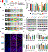
Reduction in TdTomato fluorescence established the functional efficacy and organ-targeting capability of siRNA SORT LNPs. A) B6.129TdTom/EGFP dual-reporter mice were injected twice intravenously (Day 1 and 3) with 1.5 mg kg−1 dose of siRNA SORT LNPs encapsulated with siRNA against TdTomato (siTdTom 21-mer). Major organs (lungs, liver, spleen, kidneys, heart) were harvested on Day 4 and imaged by IVIS imaging. B) IVIS images of respective organs after treatment with siRNA SORT LNPs. C) Total fluorescence intensity was quantified by drawing a ROI (region of interest) around the organ and relative change in fluorescence (compared to tdTom+ group) was calculated for each formulation. D) Western blot of total protein extract from mouse organs exhibited a reduction in TdTomato levels after treatment with respective siRNA SORT LNPs. α-Actinin was used as housekeeping control. E) Representative confocal microscopy images of organs from B6.129TdTom/EGFP mice after intravenous injections with siRNA SORT LNPs encapsulating siTdTom 21-mer. Nuclei were stained with DAPI and decrease in TdTomato fluorescence was indicative of knockdown efficacy of the LNPs. F) Elucidation of key cell types transfected by siRNA SORT LNPs by flow cytometry. On Day 4 following IVIS imaging, mouse organs (lungs, liver, spleen, and kidneys) were harvested, single cell suspensions were stained for major cell types in respective organs and analyzed by flow cytometry. Data analysis was performed with FlowJo software (v10.8.1, BD Biosciences). The % change in TdTomato intensity was quantified by normalizing TdTomato fluorescence to TdTom+ group (black bar). Groups - tdTom- (Untreated WT C57BL/6J mice, gray bar), tdTom+ (Untreated B6.129TdTom/EGFP mice, black bar), tdTomScr (B6.129TdTom/EGFP mice treated with scrambled siRNA, brown bar), and siRNA SORT LNPs treated (DOTAP-50 (red), DODAP-20 (blue), 18PA-15 (red)). Data are shown as mean ± SEM (n=3) and were analyzed in C and E by One-way ANOVA with Tukey’s multiple comparisons test (**** p < 0.0001, *** p < 0.001, ** p < 0.01, ns - non-significant).
Figure 5.
Figure 5.
Silencing endogenous Tie2 resulted in higher lung water content and an increased vascular permeability in mouse lungs. A) 6–8-week-old mice were injected intravenously twice (Day 1 and 3) with 0.5 mg kg−1 dose of DOTAP-50 LNPs containing either siScrambled or siTie2. B) Determination of lung water content after Tie2 silencing. Lungs were excised on Day 4 and wet weight was immediately recorded. The lungs were dried for 24 h at 50 °C and weighed to calculate the wet-to-dry ratio relative to untreated lungs. C) Representative images of whole lungs harvested 30 min after injection with 0.5% Evans Blue dye on Day 4. Enhancement of blue color in siTie2 group was due to leaky vasculature following Tie2 silencing. D) Quantification of Evans Blue dye extravasation in major organs after DOTAP-50 treatment. Evans Blue was extracted in formamide from pre-weighed tissue and the amount of extravasated dye (ng) per mg. of organ weight was plotted. A 2.3-fold increase in the dye content was observed in siTie2 group exclusively in the lungs when compared to both untreated and siScrambled groups. Data are shown as mean ± SEM (n=4–5) and were analyzed by Two-way ANOVA with Tukey’s multiple comparisons test (**** p < 0.0001, ** p < 0.01, ns - non-significant).

Similar articles

Cited by

References

    1. Mendes BB, Conniot J, Avital A, Yao D, Jiang X, Zhou X, Sharf-Pauker N, Xiao Y, Adir O, Liang H, Shi J, Schroeder A, Conde J, Nat Rev Methods Primers 2022, 2; - PMC - PubMed
    2. Yonezawa S, Koide H, Asai T, Adv Drug Deliv Rev 2020, 154–155, 64; - PMC - PubMed
    3. Akinc A, Maier MA, Manoharan M, Fitzgerald K, Jayaraman M, Barros S, Ansell S, Du X, Hope MJ, Madden TD, Mui BL, Semple SC, Tam YK, Ciufolini M, Witzigmann D, Kulkarni JA, van der Meel R, Cullis PR, Nat Nanotechnol 2019, 14, 1084; - PubMed
    4. d)Narasipura EA, VanKeulen-Miller R, Ma Y, Fenton OS, Bioconjug Chem 2023, 34, 1177; - PubMed
    5. Setten RL, Rossi JJ, Han SP, Nat Rev Drug Discov 2020, 19, 291; - PubMed
    6. Kulkarni JA, Witzigmann D, Chen S, Cullis PR, van der Meel R, Acc Chem Res 2019, 52, 2435. - PubMed
    1. Gomes-da-Silva LC, Fonseca NA, Moura V, Pedroso de Lima MC, Simoes S, Moreira JN, Acc Chem Res 2012, 45, 1163. - PubMed
    1. Lorenzer C, Dirin M, Winkler AM, Baumann V, Winkler J, J Control Release 2015, 203, 1; - PubMed
    2. Dong Y, Siegwart DJ, Anderson DG, Adv Drug Deliv Rev 2019, 144, 133; - PMC - PubMed
    3. Brown KM, Nair JK, Janas MM, Anglero-Rodriguez YI, Dang LTH, Peng H, Theile CS, Castellanos-Rizaldos E, Brown C, Foster D, Kurz J, Allen J, Maganti R, Li J, Matsuda S, Stricos M, Chickering T, Jung M, Wassarman K, Rollins J, Woods L, Kelin A, Guenther DC, Mobley MW, Petrulis J, McDougall R, Racie T, Bombardier J, Cha D, Agarwal S, Johnson L, Jiang Y, Lentini S, Gilbert J, Nguyen T, Chigas S, LeBlanc S, Poreci U, Kasper A, Rogers AB, Chong S, Davis W, Sutherland JE, Castoreno A, Milstein S, Schlegel MK, Zlatev I, Charisse K, Keating M, Manoharan M, Fitzgerald K, Wu JT, Maier MA, Jadhav V, Nat Biotechnol 2022, 40, 1500. - PubMed
    1. Dilliard SA, Siegwart DJ, Nat Rev Mater 2023, 8, 282; - PMC - PubMed
    2. Paunovska K, Loughrey D, Dahlman JE, Nat Rev Genet 2022, 23, 265; - PMC - PubMed
    3. Kanasty R, Dorkin JR, Vegas A, Anderson D, Nat Mater 2013, 12, 967; - PubMed
    4. Akinc A, Zumbuehl A, Goldberg M, Leshchiner ES, Busini V, Hossain N, Bacallado SA, Nguyen DN, Fuller J, Alvarez R, Borodovsky A, Borland T, Constien R, de Fougerolles A, Dorkin JR, Narayanannair Jayaprakash K, Jayaraman M, John M, Koteliansky V, Manoharan M, Nechev L, Qin J, Racie T, Raitcheva D, Rajeev KG, Sah DW, Soutschek J, Toudjarska I, Vornlocher HP, Zimmermann TS, Langer R, Anderson DG, Nat Biotechnol 2008, 26, 561. - PMC - PubMed
    1. Hou X, Zaks T, Langer R, Dong Y, Nat Rev Mater 2021, 6, 1078. - PMC - PubMed

LinkOut - more resources