Review
. 2024:49:70.

Neural pathways that compel us to scratch an itch

Affiliations
Review

Neural pathways that compel us to scratch an itch

Jagat Narayan Prajapati et al. J Biosci. 2024.

Abstract

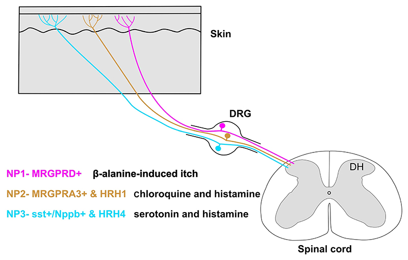
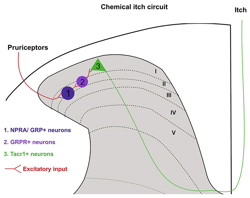
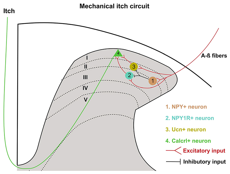
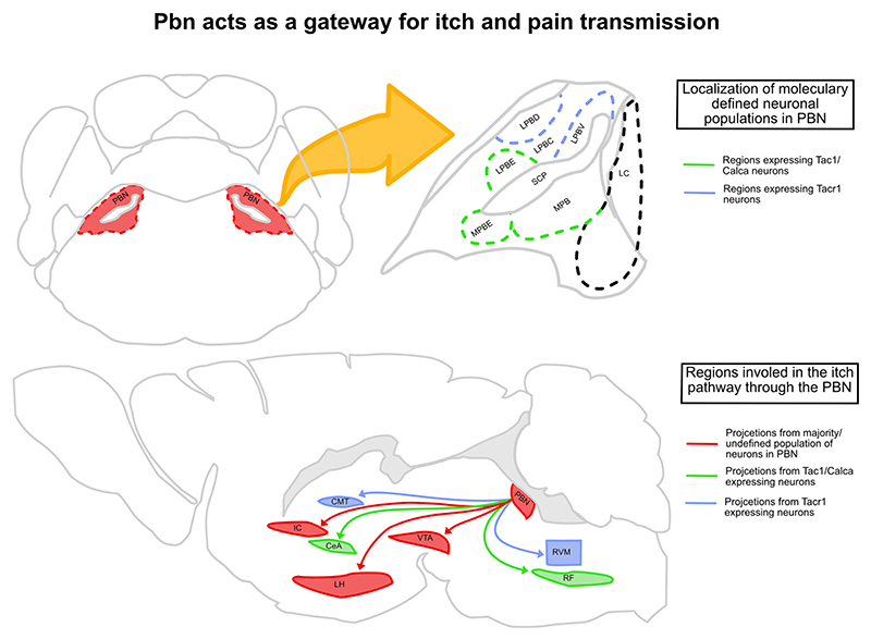
Itch is a unique sensory experience that is responded to by scratching. How pruritogens, which are mechanical and chemical stimuli with the potential to cause itch, engage specific pathways in the peripheral and central nervous system has been a topic of intense investigation over the last few years. Studies employing recently developed molecular, physiological, and behavioral techniques have delineated the dedicated mechanisms that transmit itch information to the brain. This review outlines the genetically defined and evolutionary conserved circuits for itch ranging from the skin-innervating peripheral neurons to the cortical neurons that drive scratching. Moreover, scratch suppression of itch is attributed to the concurrent activation of pain and itch pathways. Hence, we discuss the similarities between circuits driving pain and itch.


Conflict of interest statement

Conflict of interest The authors declare no conflict of interests.

Figures

Figure 1
Figure 1. The three major groups of pruriceptors in mice.
Unbiased single-cell RNA-sequencing classifies itch-sensing DRG neurons into three categories: NP1, NP2, and NP3. These neurons innervate the epidermis of the skin, and hence are positioned to sense external as well as internal pruritogens. The NP1 population of sensory neurons expresses MrgprD receptors and is involved in β-alasnine-induced itch. NP2 and NP3 both expresses the histamine receptors HRH1 and HRH4, respectively, and hence are involved in histaminergic itch. The distinction between NP2 and NP3 being, NP2 can evoke itch through chloroquine as it also expresses the MrgprA3 receptor, whereas NP3 expresses both Sst (somatostatin) and Nppb (natriuretic polypeptide b), which play an important role in the transmission of itch signals at the periphery and spinal cord levels.
Figure 2
Figure 2. The role of spinal Grp–Grpr circuitry in the itch pathway.
The Npra (natriuretic peptide receptor A), which is a receptor for neuropeptide Nppb, also expresses Grp. Hence, when Nppb-expressing pruriceptors activate dorsal horn Npra-expressing neurons during itch, Grp is released, which in turn activates Grpr interneurons. The Grpr neurons synapse onto the Tacr1 projection neurons which have been implicated in both the itch and pain pathways. This makes the Grp–Grpr circuitry important in evoking itch responses.
Figure 3
Figure 3. The pathway for mechanical itch sensation.
Both Npy1r- and Ucn-expressing interneurons promote mechanical itch but can be inhibited through Npy neurons. This indicates that transmission of mechanical itch sensation relies on the type of inputs received from the pruriceptors as they can directly synapse onto Npy1r- and Ucn-expressing interneurons. They synapse onto Calcrl-expressing neurons which project to the PBN.
Figure 4
Figure 4. The PBN acts as a gateway for itch and pain transmission.
The PBN lies laterally to the locus coeruleus (LC) and is divided into the lateral PBN and medial PBN. The majority of Tacr1-expressing cells are in the ventro-lateral (LPBV) and dorso-lateral (LPBD) part of the PBN, and majority of the Tac1- and Calca-expressing cells are in the external-lateral (LPBE), external-medial (MPBE), and medial (MPB) PBN. The Tac1/Calca cells projecting to CeA and RF (reticular formation) can mediate/affect both up- and downstream itch pathways, respectively. The Tacr1 cells projecting to CMT (centro-medial thalamus) and RVM can mediate inhibition of itch through pain. The VTA, IC (insular cortex), and LH (lateral hypothalamus) also receive strong inputs from the PBN and have been implicated in affecting various aspects of both itch and pain pathways.
Figure 5
Figure 5. Interactions of pain and itch transmission in both ascending and descending circuits.
Both pain and itch information are transmitted from the spinal cord to the CMT and PBN through the spinothalamic and spinoparabrachial pathways, respectively. The CMT directly projects to the somatosensory cortex (SSC), while the PBN projects to the IC and cingulate cortex (CC), where the anterior and posterior side of the regions code for itch and pain distinctively. They then project to the SSC and striatum. The dorsal striatum (DS) is part of an itch-specific descending pathway. The ventral periaqueductal grey (VPAG) is part of the descending pathway where the excitatory and inhibitory neurons cause an increase in itch and pain responses, respectively. Similarly, the rostral ventromedial medulla (RVM) is part of the descending pathway where the On and Off cells have a contrasting effect on pain and itch responses.
Figure 6
Figure 6. Regions involved in either mechanical or histaminergic itch that make up the itch matrix.
Mechanical itch majorly evokes activity from the claustrum, insular cortex, and regions in the basal ganglia, whereas histaminergic itch majorly evokes activity from the parabrachial nucleus and the rostral ventromedial medulla. The somatosensory cortex I/II, striatum, prefrontal cortex, centromedial thalamus, and cingulate cortex are involved in all types of itch.
Figure 7
Figure 7. The underlying circuitry for the itch–scratch reflex.
A greater sensation of itch leads to greater scratch responses where there is an increase in the force applied during scratching (increase in amplitude) and/or an increase in duration of the scratches (increase in bout duration). Scratch response is generated at the spinal cord level through a network of interneurons that receive inputs from sensory neurons and descending neurons which can code for a multitude of motor responses known as the CPG network. Activation of a specific configuration of neurons in this network leads to a scratch response as this network synapses onto the last-order interneurons and motor neurons.

Similar articles

References

    1. Acton D, Ren X, Di Costanzo S, et al. Spinal neuropeptide Y1 receptor-expressing neurons from an essential excitatory pathway for mechanical itch. Cell Rep. 2019;28:625–39.:e6. doi: 10.1016/j.celrep.2019.06.033. - DOI - PMC - PubMed
    1. Adebayo RA, Sofowora GG, Onayemi O, et al. Chloroquine-induced pruritus in malaria fever: contribution of malaria parasitaemia and the effects of prednisolone, niacin, and their combination, compared with antihistamine. Br J Clin Pharmacol. 1997;44:157–161. doi: 10.1046/j.1365-2125.1997.00612.x. - DOI - PMC - PubMed
    1. Aghahowa SE, Obianwu HO, Isah AO, et al. Chloroquine-induced pruritus. Indian J Pharm Sci. 2010;72:283–289. doi: 10.4103/0250-474X.70471. - DOI - PMC - PubMed
    1. Akiyama T, Nguyen T, Curtis E, et al. A central role for spinal dorsal horn neurons that express neurokinin-1 receptors in chronic itch. Pain. 2015;156:1240–1246. doi: 10.1097/j.pain.0000000000000172. - DOI - PMC - PubMed
    1. Akiyama T, Tominaga M, Takamori K, et al. Roles of glutamate, substance P, and gastrin-releasing peptide as spinal neurotransmitters of histaminergic and nonhistaminergic itch. Pain. 2014;155:80–92. doi: 10.1016/j.pain.2013.09.011. - DOI - PMC - PubMed