. 2024 Jun 21;9(26):28866-28878.
doi: 10.1021/acsomega.4c03413. eCollection 2024 Jul 2.

Rapid and Amplification-free Nucleic Acid Detection with DNA Substrate-Mediated Autocatalysis of CRISPR/Cas12a

Affiliations

Rapid and Amplification-free Nucleic Acid Detection with DNA Substrate-Mediated Autocatalysis of CRISPR/Cas12a

Zhongqi Zhou et al. ACS Omega. .

Abstract

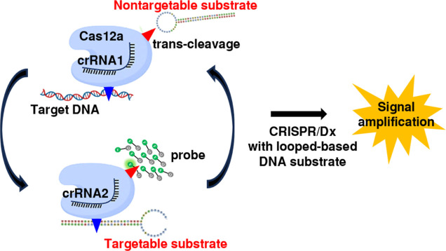
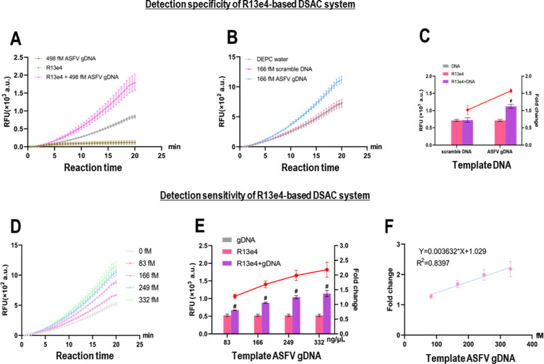
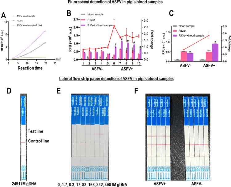
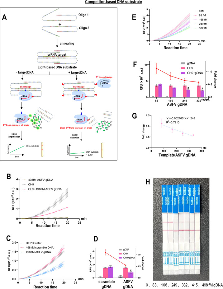
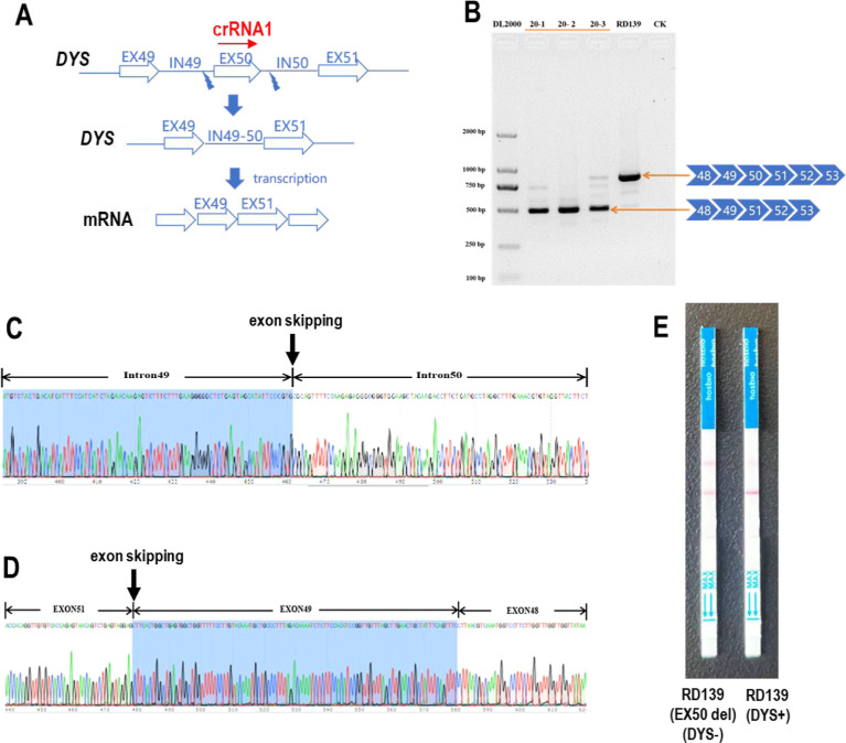
To enable rapid and accurate point-of-care DNA detection, we have developed a single-step, amplification-free nucleic acid detection platform, a DNA substrate-mediated autocatalysis of CRISPR/Cas12a (DSAC). DSAC makes use of the trans-cleavage activity of Cas12a and target template-activated DNA substrate for dual signal amplifications. DSAC employs two distinct DNA substrate types: one that enhances signal amplification and the other that negatively modulates fluorescent signals. The positive inducer utilizes nicked- or loop-based DNA substrates to activate CRISPR/Cas12a, initiating trans-cleavage activity in a positive feedback loop, ultimately amplifying the fluorescent signals. The negative modulator, which involves competitor-based DNA substrates, competes with the probes for trans-cleaving, resulting in a signal decline in the presence of target DNA. These DNA substrate-based DSAC systems were adapted to fluorescence-based and paper-based lateral flow strip detection platforms. Our DSAC system accurately detected African swine fever virus (ASFV) in swine's blood samples at femtomolar sensitivity within 20 min. In contrast to the existing amplification-free CRISPR/Dx platforms, DSAC offers a cost-effective and straightforward detection method, requiring only the addition of a rationally designed DNA oligonucleotide. Notably, a common ASFV sequence-encoded DNA substrate can be directly applied to detect human nucleic acids through a dual crRNA targeting system. Consequently, our single-step DSAC system presents an alternative point-of-care diagnostic tool for the sensitive, accurate, and timely diagnosis of viral infections with potential applicability to human disease detection.


Conflict of interest statement

The authors declare no competing financial interest.

Figures

Figure 1
Figure 1
Detection principle of a loop-based DNA substrate-mediated DSAC system. The addition of loop-based DNA substrates to the CRISPR/Cas12a complex results in a cascade amplification effect. crRNA2 is used to target the ASFV DNA sequence on the DNA substrate, while crRNA1 is used to target the desired DNA sequences such as human genomic DNA.
Figure 2
Figure 2
Functional screening of DNA substrates. Fluorescent signals (RFU, left) generated by various (A) nicked-based and (B) loop-based DNA substrates. Fold change (right) is the relative fluorescence intensity of reactions in the presence and absence of target DNA. All experiments were performed in triplicate. (C) Comparison of nicked dsDNA and nicked-based DNA substrate bearing a flap fragment. Nicked dsDNA was formed by annealing of three ssDNAs with a nick in one of the DNA strands. Fluorescent analysis shows that nicked dsDNA can be recognized by a Cas12a–crRNA complex, thereby serving as a template to activate Cas12a. Fluorescent data also confirm the ability of a flap fragment to block the recognition and binding of CRISPR/Cas12a to the R4L6 DNA substrate; therefore, the background signal is low. (D) Comparison of linear dsDNA and loop-based DNA substrates in activating Cas12a. Linear dsDNA was formed by annealing of two ssDNAs that are complementary to each other except at the 3′ end. Unmatched 3′ end has similar sequences to the loop in the DNA loop. The sequences in the loop contain a small part of the protospacer crRNA and the PAM sequences. Fluorescent analysis shows that Cas12a has better recognition in linear dsDNA than the DNA loop and therefore locates part of the protospacer crRNA, and the PAM sequences in the loop can block the Cas12a recognition and binding. P values less than 0.05 were considered statistically significant with *p < 0.05 and #p < 0.01.
Figure 3
Figure 3
Optimization of the R13e4-based DSAC system. To maximize the detection sensitivity of CRISPR/Cas12 in the DSAC system, several parameters were optimized, including the (A) reaction time of CRISPR/Cas12a and the concentrations of the (B) R13e4 DNA substrate, (C) MgCl2, (D) Cas12a, and (E) probe. Fluorescent signals (RFU, left) are generated under different experimental conditions. Fold change (right) is the relative fluorescence intensity of reactions in the presence and absence of a target DNA. All experiments were performed in triplicate. P values less than 0.05 were considered statistically significant with *p < 0.05 and #p < 0.01.
Figure 4
Figure 4
Specificity and sensitivity of the R13e4-based DSAC system. (A) R13e4 DNA substrate enhances the detection of ASFV target DNA. (B) R13e4 DNA substrate specifically detects ASFV target DNA. (C) Fluorescent signals in the presence of scrambled DNA and ASFV gDNA. (D) Dose-dependent effects of ASFV genomic DNA on R13e4-enhanced DSAC detection. (E) Using R13e4 as a DNA substrate, our DSAC system is able to distinguish target gDNA from scramble DNA at the LOD of 166fM ASFV genomic DNA. (F) Proportional of ASFV genomic DNA amount to fold change of fluorescent signals. All experiments were performed in triplicate. P values less than 0.05 were considered statistically significant with *p < 0.05 and #p < 0.01.
Figure 5
Figure 5
Fluorescent detection of ASFV in swine blood samples. (A) DNA substrate enhanced ASFV detection in swine blood samples. (B) Relative fluorescent intensity. (C) Mean relative fluorescent intensity of five samples in each group. Fold change is the relative fluorescence intensity of reactions in ASFV-positive blood samples and ASFV-negative blood samples. Lateral flow strip paper detection of ASFV in swine’s blood samples at (D) high load of ASFV genomic DNA without an added DNA substrate and (E) minimal load of ASFV genomic DNA but with an added DNA substrate. (F) DSAC system is able to distinguish blood samples with ASFV (n = 5) and without ASFV (n = 5). All experiments were performed in three replications. P values less than 0.05 were considered statistically significant with *p < 0.05 and #p < 0.01.
Figure 6
Figure 6
Target DNA detection with a competitor-based DNA substrate. (A) Scheme for a competitor-based DNA substrate DSAC system. Without the target DNA, there is no trans-cleavage activity to cleave the T-rich loop. Only Cas12a with crRNA 2 (targeting DNA substrate) is activated to bind and cleave the dsDNA on the DNA substrate. In the presence of a target DNA, Cas12a with crRNA 1 (targeting genomic DNA) is activated to bind and cleave the T-rich loops. Cas12a with crRNA 2 is also activated to bind and cleave dsDNA on the DNA substrate. The resulting two DNA fragments will compete/block the probes from trans-cleavage activity. (B) Competitor-based DNA substrates enable the detection of ASFV genomic DNA. (C,D) High specificity in detection with a competitor-based DNA substrate DSAC system. (E–G) LOD of the competitor-based DNA substrate DSAC system. (H) Application of a competitor-based DNA substrate DSAC system in lateral flow strip paper detection. All experiments were performed in triplicate. P values less than 0.05 were considered statistically significant with *p < 0.05 and #p < 0.01.
Figure 7
Figure 7
Detection of human DNA with a competitor-based DNA substrate. (A) crRNA1 target site on DYS. (B) Genomic PCR, (C) DNA sequencing, and (D) cDNA sequencing confirmation of exon-50 deletion in the DYS gene. (E) Lateral flow strip paper detection of exon-50 deletion in the DYS gene.

Similar articles

Cited by

References

    1. Stower H. Crispr-Based Diagnostics. Nat. Med. 2018, 24, 702.10.1038/s41591-018-0073-z. - DOI - PubMed
    1. Kaminski M. M.; Abudayyeh O. O.; Gootenberg J. S.; Zhang F.; Collins J. J. Crispr-Based Diagnostics. Nat. Biomed. Eng. 2021, 5, 643–656. 10.1038/s41551-021-00760-7. - DOI - PubMed
    1. Zetsche B.; Gootenberg J. S.; Abudayyeh O. O.; Slaymaker I. M.; Makarova K. S.; Essletzbichler P.; Volz S. E.; Joung J.; van der Oost J.; Regev A.; Koonin E. V.; Zhang F. Cpf1 Is a Single Rna-Guided Endonuclease of a Class 2 Crispr-Cas System. Cell 2015, 163, 759–771. 10.1016/j.cell.2015.09.038. - DOI - PMC - PubMed
    1. Chen J. S.; Ma E.; Harrington L. B.; Da Costa M.; Tian X.; Palefsky J. M.; Doudna J. A. Crispr-Cas12a Target Binding Unleashes Indiscriminate Single-Stranded Dnase Activity. Science 2018, 360, 436–439. 10.1126/science.aar6245. - DOI - PMC - PubMed
    1. Abudayyeh O. O.; Gootenberg J. S.; Essletzbichler P.; Han S.; Joung J.; Belanto J. J.; Verdine V.; Cox D. B. T.; Kellner M. J.; Regev A.; Lander E. S.; Voytas D. F.; Ting A. Y.; Zhang F. Rna Targeting with Crispr-Cas13. Nature 2017, 550, 280–284. 10.1038/nature24049. - DOI - PMC - PubMed

LinkOut - more resources