. 2024 May 16;9(26):28520-28533.
doi: 10.1021/acsomega.4c02514. eCollection 2024 Jul 2.

Lactobacillus rhamnosus 069 and Lactobacillus brevis 031: Unraveling Strain-Specific Pathways for Modulating Lipid Metabolism and Attenuating High-Fat-Diet-Induced Obesity in Mice

Affiliations

Lactobacillus rhamnosus 069 and Lactobacillus brevis 031: Unraveling Strain-Specific Pathways for Modulating Lipid Metabolism and Attenuating High-Fat-Diet-Induced Obesity in Mice

Pin-Yu Ho et al. ACS Omega. .

Abstract

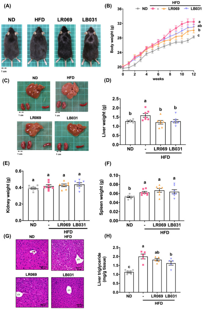
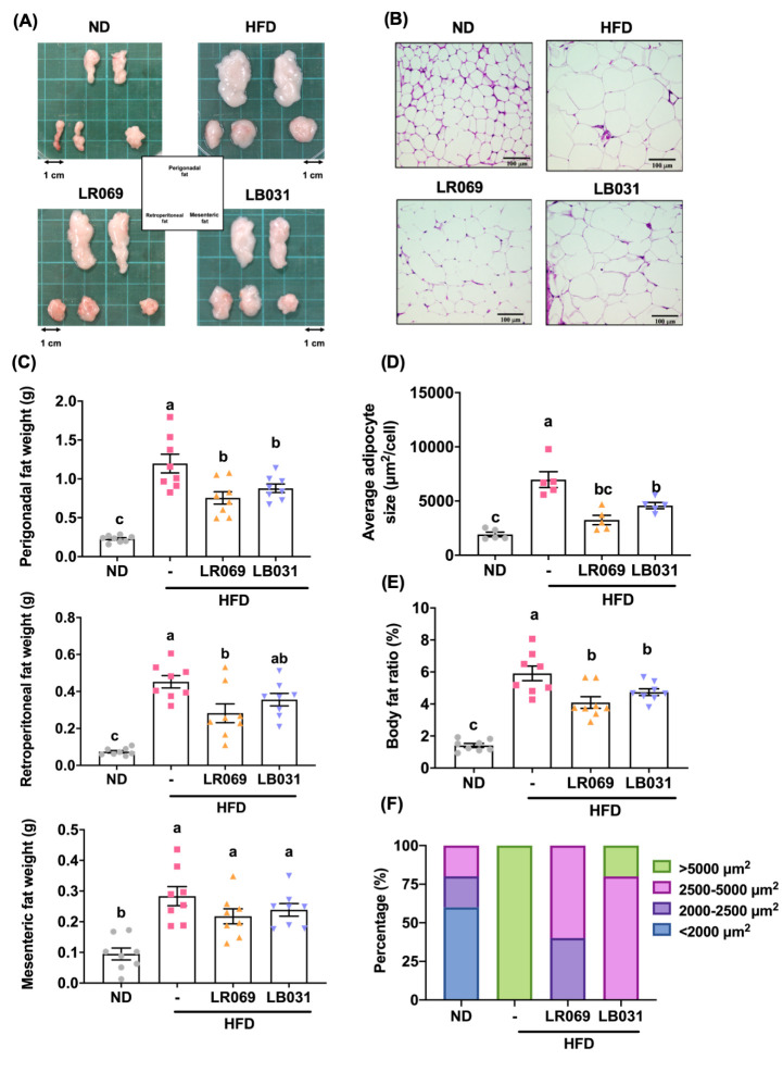
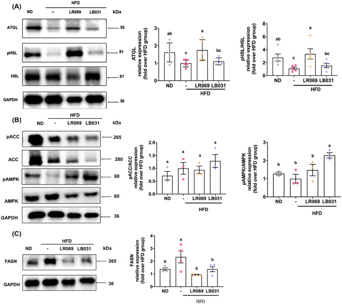
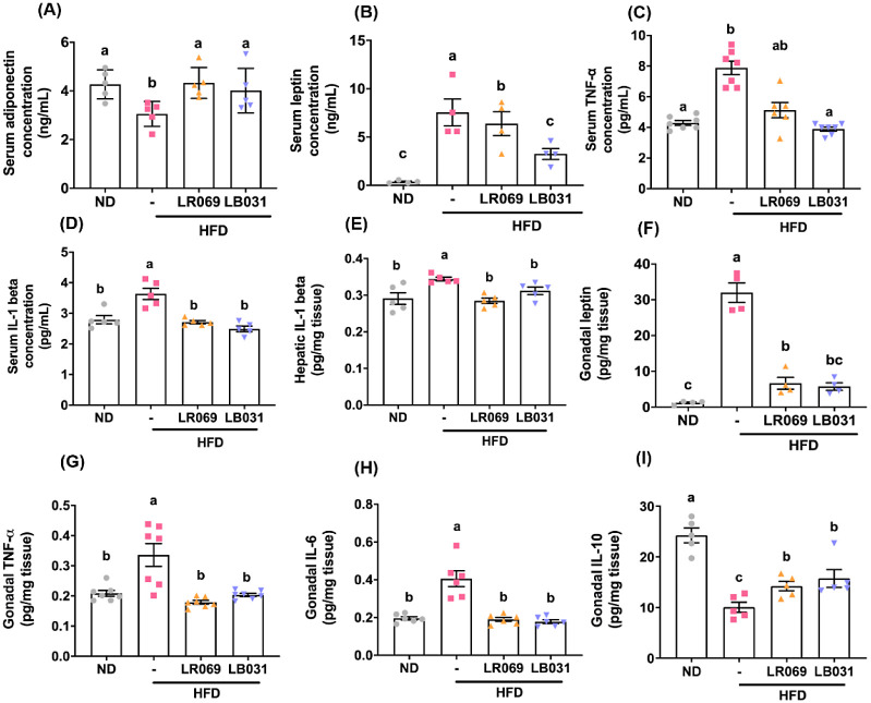
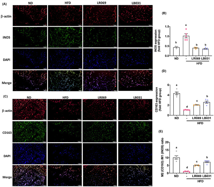
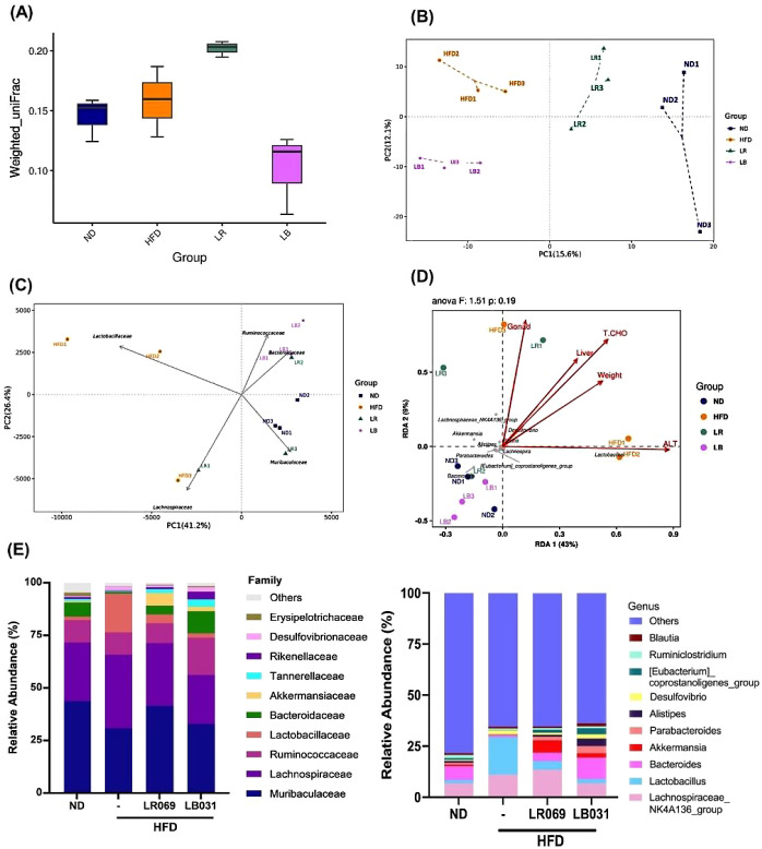
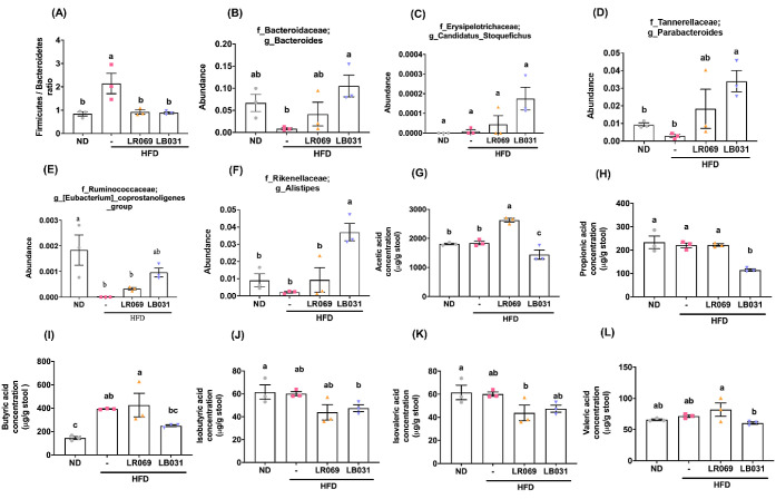
Obesity is a global health crisis, marked by excessive fat in tissues that function as immune organs, linked to microbiota dysregulation and adipose inflammation. Investigating the effects of Lactobacillus rhamnosus SG069 (LR069) and Lactobacillus brevis SG031 (LB031) on obesity and lipid metabolism, this research highlights adipose tissue's critical immune-metabolic role and the probiotics' potential against diet-induced obesity. Mice fed a high-fat diet were treated with either LR069 or LB031 for 12 weeks. Administration of LB031 boosted lipid metabolism, indicated by higher AMP-activated protein kinase (AMPK) and acetyl-CoA carboxylase (ACC) phosphorylation, and increased the M2/M1 macrophage ratio, indicating LB031's anti-inflammatory effect. Meanwhile, LR069 administration not only led to significant weight loss by enhancing lipolysis which evidenced by increased phosphorylation of hormone-sensitive lipase (HSL) and adipose triglyceride lipase (ATGL) but also elevated Akkermansia and fecal acetic acid levels, showing the gut microbiota's pivotal role in its antiobesity effects. LR069 and LB031 exhibit distinct effects on lipid metabolism and obesity, underscoring their potential for precise interventions. This research elucidates the unique impacts of these strains on metabolic health and highlights the intricate relationship between gut microbiota and obesity, advancing our knowledge of probiotics' therapeutic potential.


Conflict of interest statement

The authors declare no competing financial interest.

Figures

Figure 1
Figure 1
Effects of LR069 and LB031 on body weight, organs, H&E staining of liver, and liver triglyceride in HFD-fed mice. (A) Representative photographs of each group. (B) Weekly average body weight of each group, expressed as means ± SEM (C)−(F) Representative photographs of each group’s organs (liver, kidneys, and spleen) and organ weights. (G) Pathological assessment by H&E staining of the liver (200× magnification; length of the scale on the right = 100 μm). (H) Liver triglycerides. Data are expressed as the means ± SEM. Values with different letters (a−c) differ significantly (p < 0.05) among the compared groups.
Figure 2
Figure 2
Effect of LR069 and LB031 on adipose tissue in HFD-fed mice. (A) Representative appearances of perigonadal, mesenteric, and retroperitoneal tissues. (B) Representative image of H&E-stained perigonadal adipose tissue (200× magnification; length of the scale on the right = 100 μm). (C) Adipose tissue weights. (D) Average adipocyte size. (E) Body fat ratio (%) is calculated as the total weight of adipose tissues/body weight ×100. (F) Percentage of the adipocyte size distribution of perigonadal adipose tissue. Data are expressed as the means ± SEM. Values with different letters (a−c) differ significantly (p < 0.05) among the compared groups.
Figure 3
Figure 3
Effect of LR069 and LB031 on lipid metabolism-related proteins in the perigonadal adipose tissue in HFD-fed mice. (A) Representative Western blot images of ATGL, p-HSL, HSL, and their quantification, using GAPDH as internal control. (B) Representative Western blot images of p-ACC, ACC, p-AMPK, AMPK, and their quantification, using GAPDH as internal control. (C) Relative Western blot images of FASN and its quantification, using GAPDH as internal control. The protein bands were quantified using ImageJ software. Data are expressed as the means ± SEM. Values with different letters (a−c) differ significantly (p < 0.05) among the compared groups.
Figure 4
Figure 4
Effect of LR069 and LB031 on cytokines in HFD-fed mice. (A) Serum adiponectin concentration. (B) Serum leptin concentration. (C) Serum TNF alpha concentration. (D) Serum IL-1 beta concentration. (E) Hepatic IL-1 beta. (F) Gonadal leptin. (G) Gonadal TNF-alpha. (H) Gonadal IL-6. (I) Gonadal IL-10. Data are expressed as the means ± SEM. Values with different letters (a−c) differ significantly (p < 0.05) among the compared groups.
Figure 5
Figure 5
LR069 and LB031 regulate M1/M2 adipose tissue macrophages (ATMs) in perigonadal adipose tissue. (A) Immunofluorescence staining of β-actin (red), M1 marker iNOS (green), and nuclei (DAPI, blue) (200× magnification; length of the scale on the right = 100 μm). (C) Immunofluorescence staining of nuclei (blue), β-actin (red), and M2 marker CD163 (green) (200× magnification; length of the scale on the right = 100 μm). (B, D) Quantification of iNOS and CD163. (E) M2 (CD163)/M1 (iNOS) ratio. The immunofluorescence staining was quantified using ImageJ software. Data are expressed as the means ± SEM. Values with different letters (a−d) differ significantly (p < 0.05) among the compared groups.
Figure 6
Figure 6
Effect of LR069 and LB031, changing the gut microbiota composition in HFD-fed mice. (A) Weighted UniFrac values show the difference in β-diversity. (B) PCoA. (C) PCA plots at family level. (D) RDA analysis. The chart of the correlation between environmental factors and genus level bacterial species between groups. (E) Family and genus classification. n = 3 for each analysis, LR = LR069, LB= LB031.
Figure 7
Figure 7
Comparison of gut microbiota and fecal SCFA levels among all experimental groups in HFD-fed mice. (A) Firmicutes/Bacteroidetes ratio. Genus changes in gut microbiota: (B) Bacteroides. (C) Candidatus_Stoquefichus. (D) Parabacteroides. (E) [Eubacterium]_coprostanoigenes. (F) Alistipes. (G) Acetic acid. (H) Propionic acid. (I) Butyric acid. (J) Isobutyric acid. (K) Isovaleric acid. (L) Valeric acid. Data are expressed as the means ± SEM. Values with different letters (a−c) differ significantly (p < 0.05) among the compared groups.

Similar articles

Cited by

References

    1. Jo J.; Gavrilova O.; Pack S.; Jou W.; Mullen S.; Sumner A. E.; Cushman S. W.; Periwal V. Hypertrophy and/or hyperplasia: Dynamics of adipose tissue growth. PLoS Comput. Biol. 2009, 5 (3), e100032410.1371/journal.pcbi.1000324. - DOI - PMC - PubMed
    1. Drozdz D.; Alvarez-Pitti J.; Wójcik M.; Borghi C.; Gabbianelli R.; Mazur A.; Herceg-Čavrak V.; Lopez-Valcarcel B. G.; Brzeziński M.; Lurbe E.; et al. Obesity and cardiometabolic risk factors: From childhood to adulthood. Nutrients 2021, 13 (11), 4176.10.3390/nu13114176. - DOI - PMC - PubMed
    1. Tiwari A.; Balasundaram P.; Public Health Considerations Regarding Obesity; StatPearls Publishing: Treasure Island, FL, 2023. - PubMed
    1. Sahu B.; Tikoo O.; Pati B.; Senapati U.; Bal N. C.. Role of distinct fat depots in metabolic regulation and pathological implications. Rev. Physiol. Biochem. Pharmacol. 2022, 186, 135–176.. 10.1007/112_2022_73. - DOI - PubMed
    1. Palhinha L.; Liechocki S.; Hottz E. D.; Pereira J. A. D. S.; de Almeida C. J.; Moraes-Vieira P. M. M.; Bozza P. T.; Maya-Monteiro C. M. Leptin induces proadipogenic and proinflammatory signaling in adipocytes. Front. Endocrinol. 2019, 10, 841.10.3389/fendo.2019.00841. - DOI - PMC - PubMed

LinkOut - more resources