

Prominent response to savolitinib monotherapy in high-grade fetal adenocarcinoma with MET amplification and concurrent brain metastasis: a case report
- PMID: 38973955
- PMCID: PMC11225042
- DOI: 10.21037/tlcr-24-124
Prominent response to savolitinib monotherapy in high-grade fetal adenocarcinoma with MET amplification and concurrent brain metastasis: a case report
Abstract
Background: Mesenchymal-epithelial transition (MET) represents a potential therapeutic target in various cancers, with amplification of the MET gene identified in a subset of patients with pulmonary adenocarcinomas. However, MET gene amplification is rarely observed in high-grade fetal adenocarcinoma (H-FLAC).
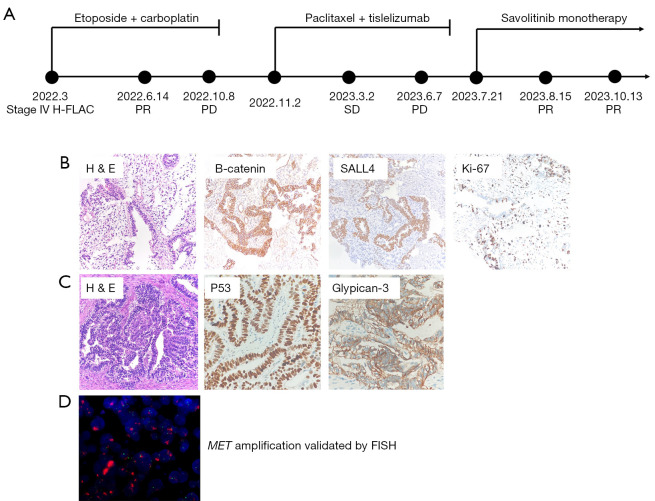
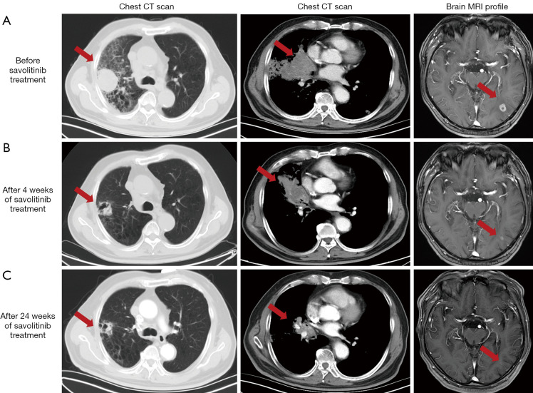
Case description: Here we present a novel case of a patient diagnosed with stage IV H-FLAC harboring MET amplifications and treated with savolitinib. The 69-year-old male patient, who presented with a primary complaint of cough and white sputum, had a history of hypertension for over 10 years and a 45-year smoking history. The patient received savolitinib monotherapy treatment due to brain metastases. Despite the omission of radiotherapy for asymptomatic brain metastases, a notable response to savolitinib therapy was observed, with a partial response (PR) achieved after 4 weeks and a reduction in the brain tumor. At the time of the submission of this report, the patient received over 24 weeks of savolitinib treatment, and was maintained PR. The patient was still undergoing treatment. This highlights the potential clinical benefits of targeted therapy against MET amplification in H-FLAC.
Conclusions: H-FLAC harboring MET amplification and brain metastasis is rare. Treatment with savolitinib monotherapy resulted in a PR, providing preliminary insights to the efficacy of savolitinib for H-FLAC with MET amplification.
Keywords: Case report; high-grade fetal adenocarcinoma (H-FLAC); mesenchymal-epithelial transition (MET); savolitinib.
2024 Translational Lung Cancer Research. All rights reserved.
Conflict of interest statement
Conflicts of Interest: All authors have completed the ICMJE uniform disclosure form (available at https://tlcr.amegroups.com/article/view/10.21037/tlcr-24-124/coif). The authors have no conflicts of interest to declare.
Figures
References
-
- Nakatani Y, Kitamura H, Inayama Y, et al. Pulmonary adenocarcinomas of the fetal lung type: a clinicopathologic study indicating differences in histology, epidemiology, and natural history of low-grade and high-grade forms. Am J Surg Pathol 1998;22:399-411. 10.1097/00000478-199804000-00003 - DOI - PubMed
-
- Suzuki M, Yazawa T, Ota S, et al. High-grade fetal adenocarcinoma of the lung is a tumour with a fetal phenotype that shows diverse differentiation, including high-grade neuroendocrine carcinoma: a clinicopathological, immunohistochemical and mutational study of 20 cases. Histopathology 2015;67:806-16. 10.1111/his.12711 - DOI - PubMed
Publication types
LinkOut - more resources
Full Text Sources
Research Materials
Miscellaneous