. 2024 Jun 12:40:212-226.
doi: 10.1016/j.bioactmat.2024.06.007. eCollection 2024 Oct.

Poly(p-coumaric acid) nanoparticles alleviate temporomandibular joint osteoarthritis by inhibiting chondrocyte ferroptosis

Affiliations

Poly(p-coumaric acid) nanoparticles alleviate temporomandibular joint osteoarthritis by inhibiting chondrocyte ferroptosis

Jiaxin Guo et al. Bioact Mater. .

Abstract

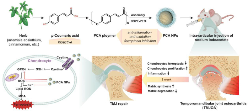
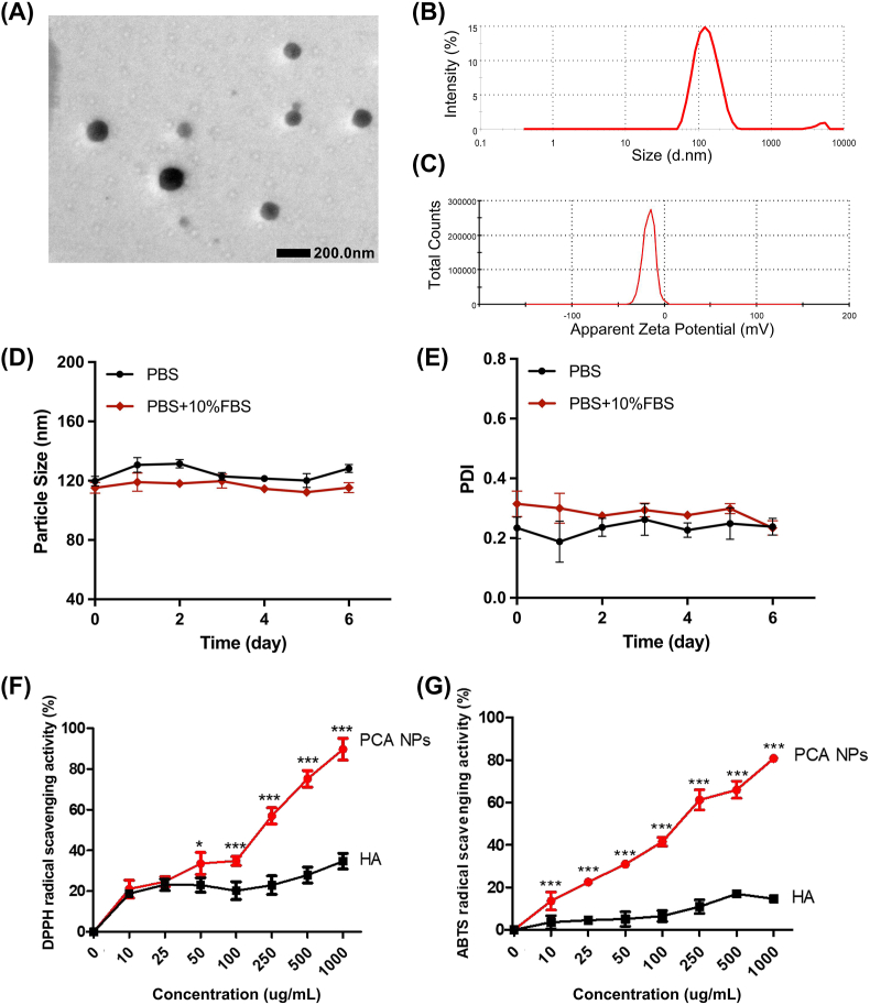
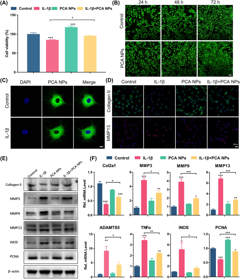
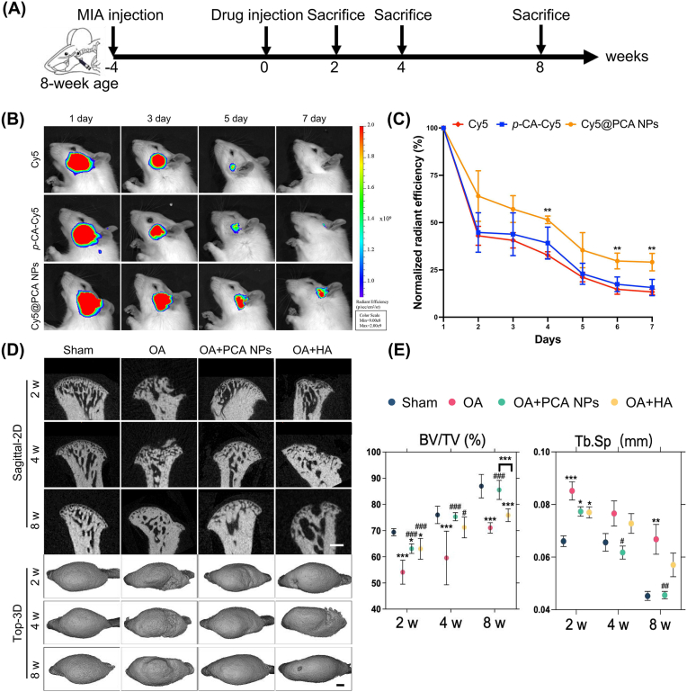
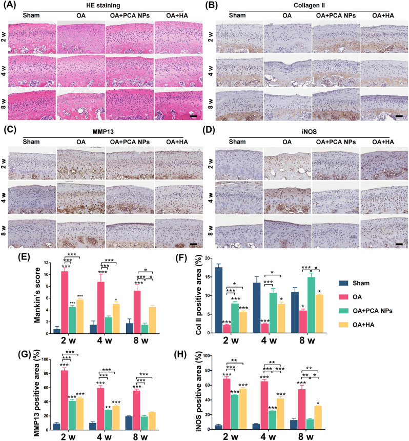
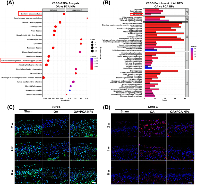
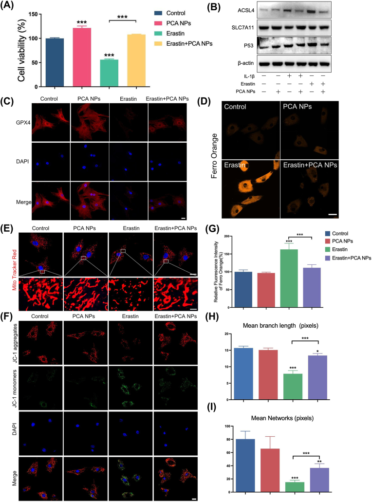
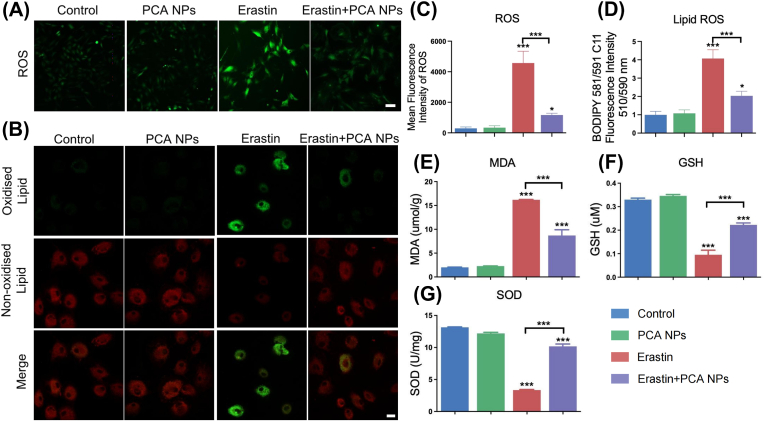
Oxidative stress and inflammation are key drivers of osteoarthritis (OA) pathogenesis and disease progression. Herein we report the synthesis of poly(p-coumaric) nanoparticles (PCA NPs) from p-courmaic acid (p-CA), a naturally occurring phytophenolic acid, to be a multifunctional and drug-free therapeutic for temporomandibular joint osteoarthritis (TMJOA). Compared to hyaluronic acid (HA) that is clinically given as viscosupplementation, PCA NPs exhibited long-term efficacy, superior anti-oxidant and anti-inflammatory properties in alleviating TMJOA and repairing the TMJ cartilage and subchondral bone in a rat model of TMJOA. Notably, TMJ repair mediated by PCA NPs could be attributed to their anti-oxidant and anti-inflammatory properties in enhancing cell proliferation and matrix synthesis, while reducing inflammation, oxidative stress, matrix degradation, and chondrocyte ferroptosis. Overall, our study demonstrates a multifunctional nanoparticle, synthesized from natural p-coumaric acid, that is stable and possess potent antioxidant, anti-inflammatory properties and ferroptosis inhibition, beneficial for treatment of TMJOA.

Keywords: Cartilage; Ferroptosis; Oxidative pressure; Poly(p-coumaric acid) nanoparticles; Temporomandibular joint osteoarthritis.


Conflict of interest statement

The authors declare that they have no known competing financial interests or personal relationships that could have appeared to influence the work reported in this paper.

Figures

Image 1
Graphical abstract
Scheme 1
Scheme 1
The mechanism of PCA NPs contribute to the mitigation of TMJOA progression by lowering matrix degradation, increasing proliferation and matrix synthesis, attenuating oxidation and inflammation, and inhibiting chondrocyte ferroptosis.
Fig. 1
Fig. 1
Characterization of PCA NPs. (A) Typical TEM photograph showcasing PCA NPs. (B) Dimension distribution of PCA NPs as determined by DLS. (C) Measurement of the zeta potential distribution for PCA NPs. (D and E) Assessment of PCA NPs stability in PBS and PBS supplemented with 10 % FBS. (F and G) Comparative analysis of in vitro antioxidant efficacy of PCA NPs against HA. *P < 0.05, **P < 0.01, ***P < 0.001 compared to HA.
Fig. 2
Fig. 2
Chondroprotective effects of PCA NPs on chondrocytes stimulated under IL-1β-induced inflammation in vitro. (A) Effects of PCA NPs on viability of IL-1β-treated chondrocytes. (B) Live/dead staining of chondrocytes treated with 75 μg/mL PCA NPs. Scale bar: 100 μm. (C) Internalization of C6-labbled PCA NPs by chondrocytes, with or without IL-1β stimulation for 24 h. Scale bar: 10 μm. (D) Representative immunofluorescence pictures showing Collagen II and MMP13 in chondrocytes. Scale bar: 50 μm. (E) Western blot analysis of anabolic markers (Collagen II and PCNA), and catabolic markers (MMP3, MMP9, MMP13 and iNOS). (F) Gene expression analysis of anabolic markers (Col2a and PCNA) and catabolic markers (MMP3, MMP9, MMP13, ADAMTS5, TNFα and iNOS). *P < 0.05, **P < 0.01, and ***P < 0.001 compared to the control group.
Fig. 3
Fig. 3
PCA NPs alleviate subchondral bone destruction and promote TMJ repair in TMJOA. (A) Depiction of the in vivo experimental procedure. (B and C) Representative fluorescence pictures (B) and quantitative analysis of normalized flsorescence radiant efficiency within the rat TMJ (C) at various time points after removing the hair and intra-articular injection of free Cy5, p-CA-Cy5 and Cy5@PCA NPs. **P < 0.01 compared to the free Cy5 group. (D and E) Rat TMJs were collected for micro-CT examination at intervals of 2, 4, and 8 weeks. (D) Sagittal and top views of the condyles. Scale bar: 500 μm. (E) Analysis of BV/TV and Tb.Sp. *P < 0.05, **P < 0.01, ***P < 0.001 compare to the sham rats; #P < 0.05, ##P < 0.01, ###P < 0.001 in contrast to the OA rats.
Fig. 4
Fig. 4
PCA NPs alleviated condylar cartilage degeneration in TMJOA. (A–D) Histological staining by (A) hematoxylin and eosin (HE) and immunohistochemical staining for (B) Collagen II, (C) MMP13, and (D) iNOS at 2, 4 and 8 weeks following treatment. Scale bar: 50 μm. (E) Assessment of Mankin's score. (F–H) Quantitative analysis of (F) Collagen II+ area, (G) iNOS+ cells, and (H) MMP13+ cells in the condylar cartilage over 2, 4, and 8 weeks. *P < 0.05, **P < 0.01, ***P < 0.001 compared to the sham group at corresponding time points.
Fig. 5
Fig. 5
PCA NPs inhibited chondrocyte ferroptosis in rat TMJOA. (A and B) Transcriptomic analysis of TMJ condylar cartilage tissue. (A) Results from KEGG GSEA analysis comparing the OA and PCA NPs groups. (B) Results from KEGG enrichment of all DEG analysis comparing the OA and PCA NPs groups. (C and D) Exemplary immunofluorescence staining pictures for GPX4 and ACSL4. Scale bar: 40 μm.
Fig. 6
Fig. 6
PCA NPs alleviated Fe2+ overload and mitochondrial dysfunction in chondrocytes stimulated with erastin. (A) Impact of PCA NPs on IL-1β-stimulated chondrocyte activity. (B) Western blotting was used to assess ACLS4, SLC7A11, and P53 protein expression in chondrocytes. (C) Immunofluorescence staining for GPX4 in chondrocytes. Scale bar: 20 μm. (D and G) FerroOrange flsorescence probe staining (D) with its quantitative analysis (G) for Fe2+ in chondrocytes. Scale bar: 20 μm. (E, H and I) MitoTracker Red staining (E) along with quantitative evaluations of average mitochondrial branch length (H) and network (I) in chondrocytes. Scale bar: 20 μm. (The enlarge scale bar: 2 μm). (F) JC-1 staining for assessing mitochondrial membrane potential of chondrocytes. Scale bar: 20 μm *P < 0.05, **P < 0.01, ***P < 0.001 in comparison to the control group.
Fig. 7
Fig. 7
PCA NPs inhibited chondrocyte lipid peroxidation induced by erastin. (A–D) Representative fluorescence images for cellular ROS (A) and lipid ROS (B), along with their quantitative assessment via flow cytometry (C and D). Scale bar: 20 μm. (E–G) Quantitative determination of MDA (E), GSH (F), and SOD (G) Levels in cells treated with PCA NPs and stimulated with erastin. *P < 0.05, **P < 0.01, ***P < 0.001 in contrast to the control.

Similar articles

Cited by

References

    1. Quicke J.G., Conaghan P.G., Corp N., Peat G. Osteoarthritis year in review 2021: epidemiology & therapy. Osteoarthritis Cartilage. 2022;30(2):196–206. - PubMed
    1. Yao Q., Wu X., Tao C., Gong W., Chen M., Qu M., Zhong Y., He T., Chen S., Xiao G. Osteoarthritis: pathogenic signaling pathways and therapeutic targets. Signal Transduct. Targeted Ther. 2023;8(1):56. - PMC - PubMed
    1. Hill A., Duran J., Purcell P. Lubricin protects the temporomandibular joint surfaces from degeneration. PLoS One. 2014;9(9) - PMC - PubMed
    1. Wang X.D., Zhang J.N., Gan Y.H., Zhou Y.H. Current understanding of pathogenesis and treatment of TMJ osteoarthritis. J. Dent. Res. 2015;94(5):666–673. - PubMed
    1. Barry F., Chai F., Chijcheapaza-Flores H., Garcia-Fernandez M.J., Blanchemain N., Nicot R. Systematic review of studies on drug-delivery systems for management of temporomandibular-joint osteoarthritis. J Stomatol Oral Maxillofac Surg. 2022;123(5):e336–e341. - PubMed

LinkOut - more resources