

Ovarian cancer ascites proteomic profile reflects metabolic changes during disease progression
- PMID: 38974022
- PMCID: PMC11225207
- DOI: 10.1016/j.bbrep.2024.101755
Ovarian cancer ascites proteomic profile reflects metabolic changes during disease progression
Abstract
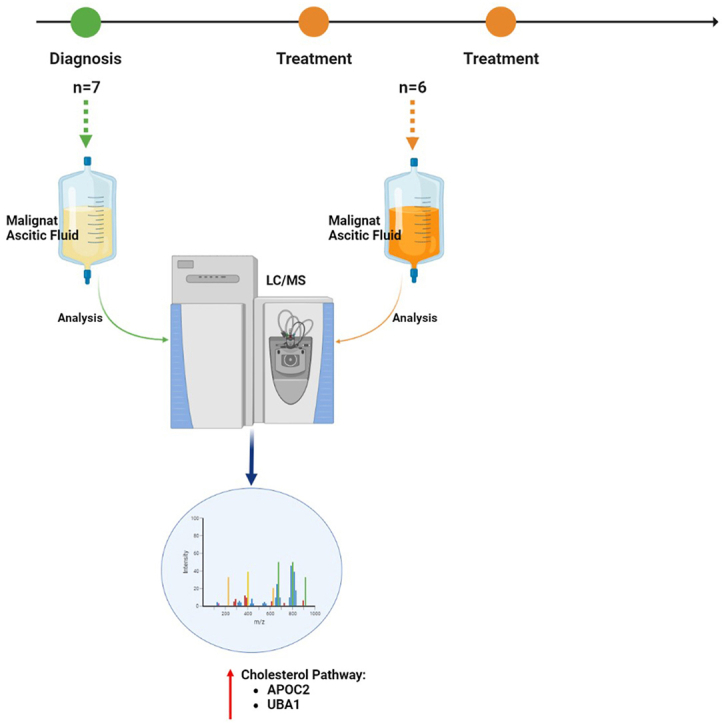
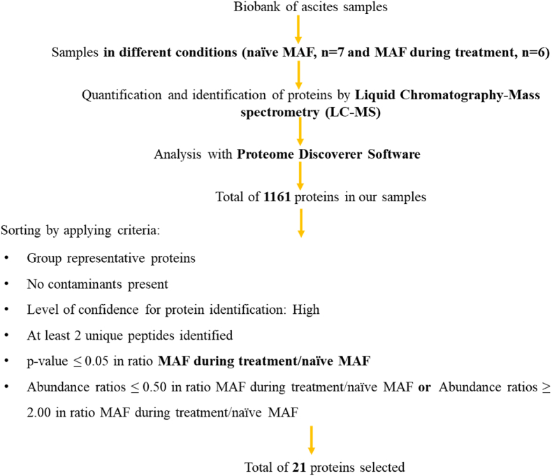
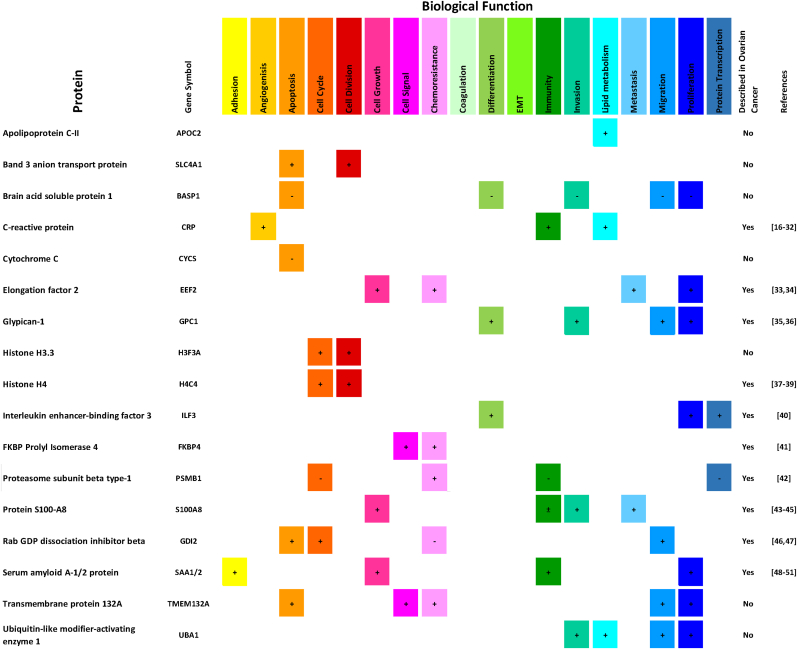
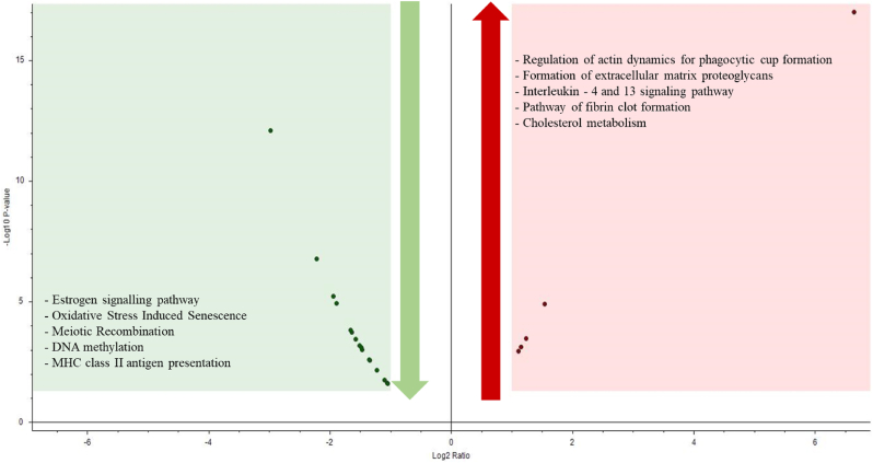
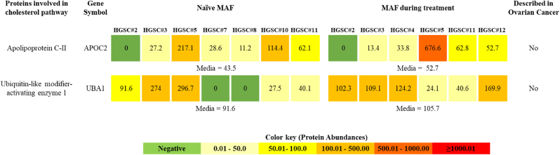
Ovarian cancer (OC) patients develop ascites, an accumulation of ascitic fluid in the peritoneal cavity anda sign of tumour dissemination within the peritoneal cavity. This body fluid is under-researched, mainly regarding the ascites formed during tumour progression that have no diagnostic value and, therefore, are discarded. We performed a discovery proteomics study to identify new biomarkers in the ascites supernatant of OC patients. In this preliminary study, we analyzed a small amount of OC ascites to highlight the importance of not discarding such biological material during treatment, which could be valuable for OC management. Our findings reveal that OC malignant ascitic fluid (MAF) displays a proliferative environment that promotes the growth of OC cells that shift the metabolic pathway using alternative sources of nutrients, such as the cholesterol pathway. Also, OC ascites drained from patients during treatment showed an immunosuppressive environment, with up-regulation of proteins from the signaling pathways of IL-4 and IL-13 and down-regulation from the MHC-II. This preliminary study pinpointed a new protein (Transmembrane Protein 132A) in the OC context that deserves to be better explored in a more extensive cohort of patients' samples. The proteomic profile of MAF from OC patients provides a unique insight into the metabolic kinetics of cancer cells during disease progression, and this information can be used to develop more effective treatment strategies.
Keywords: High-grade serous carcinoma; Malignant ascitic fluid; Metabolic pathways; Proteomics; Tumor microenvironment.
© 2024 The Authors. Published by Elsevier B.V.
Conflict of interest statement
The authors declare that they have no known competing financial interests or personal relationships that could have appeared to influence the work reported in this paper.
Figures
References
-
- Shender V.O., Pavlyukov M.S., Ziganshin R.H., Arapidi G.P., Kovalchuk S.I., Anikanov N.A., Altukhov I.A., Alexeev D.G., Butenko I.O., Shavarda A.L., et al. Proteome–metabolome profiling of ovarian cancer ascites reveals novel components involved in intercellular communication. Mol. Cell. Proteomics. 2014;13:3558–3571. doi: 10.1074/mcp.M114.041194. - DOI - PMC - PubMed
LinkOut - more resources
Full Text Sources
Research Materials
