

The Prevalence of the Middle Clinoid Process: A Cross-Sectional Comparative Study in Patients with and without Pathology of the Sella Turcica
- PMID: 38974427
- PMCID: PMC11226267
- DOI: 10.1055/s-0044-1787054
The Prevalence of the Middle Clinoid Process: A Cross-Sectional Comparative Study in Patients with and without Pathology of the Sella Turcica
Abstract
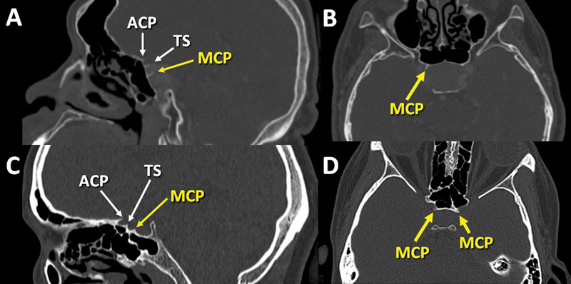
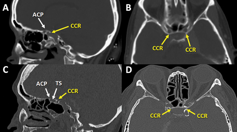
Background The middle clinoid process (MCP), particularly caroticoclinoid ring (CCR) type of the MCP, is an important part of the sphenoid bone for skull base surgery. Previous studies have shown a wide range of MCP prevalence affected by various factors. However, no study has investigated the association between the MCP and the presence of sellar lesions. Objectives The main aim of this study was to evaluate the prevalence of the MCP in the Thai population and factors associated with its presence. Materials and Methods We conducted a cross-sectional study on 400 sides from 200 patients (100 with and 100 without sellar lesions) using cranial computerized tomography scans. Demographic data and MCP characteristics were collected. The association between individual variables and the presence of the MCP was determined by univariate and multivariate analysis. Results The MCP was identified in 168 of 400 sides (42%). Patients with sellar lesions had a significantly lower prevalence of the MCP compared with normal controls (29.5% versus 54.5%, p < 0.001). Of all MCP only 6% were the CCR type. Univariate and multivariate analysis showed that the absence of the sellar lesion was the only factor significantly associated with presence of the MCP (odds ratio: 2.86; 95% confidence interval: 1.90-4.32; p < 0.001). Conclusion The prevalence of the MCP was relatively high in the Thai population, while the prevalence of the CCR was relatively low compared with previous studies. The absence of sellar lesions was the only factor associated with the presence of the MCP.
Keywords: caroticoclinoid ring; cranial computerized tomography; middle clinoid process; prevalence; skull base surgery.
Asian Congress of Neurological Surgeons. This is an open access article published by Thieme under the terms of the Creative Commons Attribution-NonDerivative-NonCommercial License, permitting copying and reproduction so long as the original work is given appropriate credit. Contents may not be used for commercial purposes, or adapted, remixed, transformed or built upon. ( https://creativecommons.org/licenses/by-nc-nd/4.0/ ).
Conflict of interest statement
Conflict of Interest None declared.
Figures

References
-
- Inoue T, Rhoton A L, Jr, Theele D, Barry M E. Surgical approaches to the cavernous sinus: a microsurgical study. Neurosurgery. 1990;26(06):903–932. - PubMed
-
- Fernandez-Miranda J C, Tormenti M, Latorre F, Gardner P, Snyderman C.Endoscopic endonasal middle clinoidectomy: anatomic, radiological, and technical note Neurosurgery 201271(2, suppl Operative):ons233–ons239. - PubMed
-
- Sharma A, Rieth G E, Tanenbaum J E et al. A morphometric survey of the parasellar region in more than 2700 skulls: emphasis on the middle clinoid process variants and implications in endoscopic and microsurgical approaches. J Neurosurg. 2018;129(01):60–70. - PubMed
-
- Miller C, Chamoun R, Beahm D. Morphometric analysis of the middle clinoid process using maxillofacial computed tomography scans. Oper Neurosurg (Hagerstown) 2017;13(01):124–130. - PubMed
-
- Keyes J EL. Observations on four thousand optic foramina in human skulls of known origin. Arch Ophthalmol. 1935;13(04):538–568.
LinkOut - more resources
Full Text Sources
Miscellaneous