

Predictors for the Differentiation between Glioblastoma, Primary Central Nervous System Lymphoma, and Metastasis in Patients with a Solitary Enhancing Intracranial Mass
- PMID: 38974428
- PMCID: PMC11226298
- DOI: 10.1055/s-0044-1787051
Predictors for the Differentiation between Glioblastoma, Primary Central Nervous System Lymphoma, and Metastasis in Patients with a Solitary Enhancing Intracranial Mass
Abstract
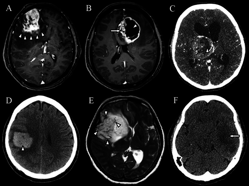
Introduction Differentiation between glioblastoma (GBM), primary central nervous system lymphoma (PCNSL), and metastasis is important in decision-making before surgery. However, these malignant brain tumors have overlapping features. This study aimed to identify predictors differentiating between GBM, PCNSL, and metastasis. Materials and Methods Patients with a solitary intracranial enhancing tumor and a histopathological diagnosis of GBM, PCNSL, or metastasis were investigated. All patients with intracranial lymphoma had PCNSL without extracranial involvement. Demographic, clinical, and radiographic data were analyzed to determine their associations with the tumor types. Results The predictors associated with GBM were functional impairment ( p = 0.001), large tumor size ( p < 0.001), irregular tumor margin ( p < 0.001), heterogeneous contrast enhancement ( p < 0.001), central necrosis ( p < 0.001), intratumoral hemorrhage ( p = 0.018), abnormal flow void ( p < 0.001), and hypodensity component on noncontrast cranial computed tomography (CT) scan ( p < 0.001). The predictors associated with PCNSL comprised functional impairment ( p = 0.005), deep-seated tumor location ( p = 0.006), homogeneous contrast enhancement ( p < 0.001), absence of cystic appearance ( p = 0.008), presence of hypointensity component on precontrast cranial T1-weighted magnetic resonance imaging (MRI; p = 0.027), and presence of isodensity component on noncontrast cranial CT ( p < 0.008). Finally, the predictors for metastasis were an infratentorial ( p < 0.001) or extra-axial tumor location ( p = 0.035), smooth tumor margin ( p < 0.001), and presence of isointensity component on cranial fluid-attenuated inversion recovery MRI ( p = 0.047). Conclusion These predictors may be used to differentiate between GBM, PCNSL, and metastasis, and they are useful in clinical management.
Keywords: brain metastasis; differentiation; glioblastoma; predictor; primary central nervous system lymphoma (PCNSL).
Asian Congress of Neurological Surgeons. This is an open access article published by Thieme under the terms of the Creative Commons Attribution-NonDerivative-NonCommercial License, permitting copying and reproduction so long as the original work is given appropriate credit. Contents may not be used for commercial purposes, or adapted, remixed, transformed or built upon. ( https://creativecommons.org/licenses/by-nc-nd/4.0/ ).
Conflict of interest statement
Conflict of Interest None declared.
Figures


Similar articles
-
Can morphological MRI differentiate between primary central nervous system lymphoma and glioblastoma?Cancer Imaging. 2016 Nov 29;16(1):40. doi: 10.1186/s40644-016-0098-9. Cancer Imaging. 2016. PMID: 27894359 Free PMC article.
-
Primary central nervous system lymphoma and glioblastoma differentiation based on conventional magnetic resonance imaging by high-throughput SIFT features.Int J Neurosci. 2018 Jul;128(7):608-618. doi: 10.1080/00207454.2017.1408613. Epub 2017 Dec 12. Int J Neurosci. 2018. PMID: 29183170
-
Primary central nervous system lymphoma and atypical glioblastoma: differentiation using the initial area under the curve derived from dynamic contrast-enhanced MR and the apparent diffusion coefficient.Eur Radiol. 2017 Apr;27(4):1344-1351. doi: 10.1007/s00330-016-4484-2. Epub 2016 Jul 19. Eur Radiol. 2017. PMID: 27436023
-
Machine learning applications for the differentiation of primary central nervous system lymphoma from glioblastoma on imaging: a systematic review and meta-analysis.Neurosurg Focus. 2018 Nov 1;45(5):E5. doi: 10.3171/2018.8.FOCUS18325. Neurosurg Focus. 2018. PMID: 30453459
-
Classifying primary central nervous system lymphoma from glioblastoma using deep learning and radiomics based machine learning approach - a systematic review and meta-analysis.Front Oncol. 2022 Oct 3;12:884173. doi: 10.3389/fonc.2022.884173. eCollection 2022. Front Oncol. 2022. PMID: 36263203 Free PMC article.
References
-
- Purandare N C, Puranik A, Shah S et al.Common malignant brain tumors: can 18F-FDG PET/CT aid in differentiation? Nucl Med Commun. 2017;38(12):1109–1116. - PubMed
-
- Ma J H, Kim H S, Rim N J, Kim S H, Cho K G. Differentiation among glioblastoma multiforme, solitary metastatic tumor, and lymphoma using whole-tumor histogram analysis of the normalized cerebral blood volume in enhancing and perienhancing lesions. AJNR Am J Neuroradiol. 2010;31(09):1699–1706. - PMC - PubMed
-
- Ling S M, Roach M, III, Larson D A, Wara W M. Radiotherapy of primary central nervous system lymphoma in patients with and without human immunodeficiency virus. Ten years of treatment experience at the University of California San Francisco. Cancer. 1994;73(10):2570–2582. - PubMed
-
- Reni M, Ferreri A J, Garancini M P, Villa E. Therapeutic management of primary central nervous system lymphoma in immunocompetent patients: results of a critical review of the literature. Ann Oncol. 1997;8(03):227–234. - PubMed
LinkOut - more resources
Full Text Sources