

Eosinophilic granuloma of the cervical spine in a young adult: A rare case report
- PMID: 38974539
- PMCID: PMC11225499
- DOI: 10.25259/SNI_262_2024
Eosinophilic granuloma of the cervical spine in a young adult: A rare case report
Abstract
Background: Spinal eosinophilic granulomas (EG) are rare tumors, mostly reported in the pediatric age group. They constitute <1% of primary bone neoplasms, and cervical spine involvement is uncommon.
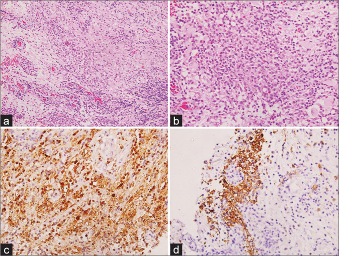
Case description: A 20-year-old male presented with neck pain for a 4-month duration. Six years previously, he had received six cycles of vinblastine for biopsy-proven histiocytosis of an axillary lymph node; this resulted in incomplete remission. Present magnetic resonance/computed tomography (CT) imaging revealed a lytic C2 body lesion with atlantoaxial instability. When the CT-guided biopsy was suggestive of EG, he was managed with definitive surgery and adjuvant radiotherapy.
Conclusion: Cervical spine EG is rare in adults. CT-guided biopsy should confirm the diagnosis and should be followed by definitive surgery and adjuvant radiotherapy.
Keywords: Adults; Cervical spine; Eosinophilic granuloma; Spine.
Copyright: © 2024 Surgical Neurology International.
Conflict of interest statement
There are no conflicts of interest.
Figures

References
-
- Bertram C, Madert J, Eggers C. Eosinophilic granuloma of the cervical spine. Spine (Phila Pa 1976) 2002;27:1408–13. - PubMed
-
- Huang W, Yang X, Cao D, Xiao J, Yang M, Feng D, et al. Eosinophilic granuloma of spine in adults: A report of 30 cases and outcome. Acta Neurochir (Wien) 2010;152:1129–37. - PubMed
-
- Huang WD, Yang XH, Wu ZP, Huang Q, Xiao JR, Yang MS. Langerhans cell histiocytosis of spine: A comparative study of clinical, imaging features, and diagnosis in children, adolescents, and adults. Spine J. 2013;13:1108–17. - PubMed
-
- Jiang L, Liu ZJ, Liu XG, Zhong WQ, Ma QJ, Wei F, et al. Langerhans cell histiocytosis of the cervical spine. A single Chinese institution experience with thirty cases. Spine (Phila Pa 1976) 2010;35:E8–15. - PubMed
Publication types
LinkOut - more resources
Full Text Sources
Miscellaneous