

Real-time display of intracranial subdural electrodes and the brain surface during an electrode implantation procedure using permeable film
- PMID: 38974543
- PMCID: PMC11225510
- DOI: 10.25259/SNI_74_2024
Real-time display of intracranial subdural electrodes and the brain surface during an electrode implantation procedure using permeable film
Abstract
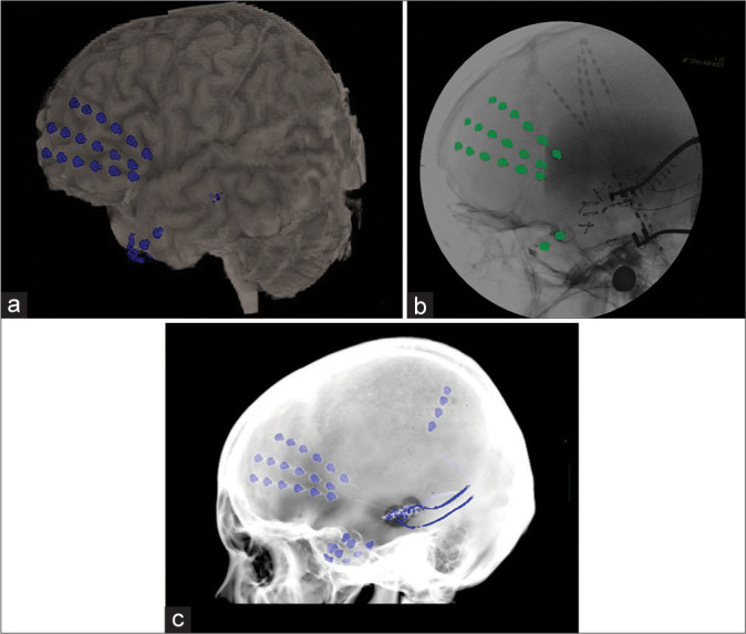
Background: Subdural electrode (SDE) implantation is an important method of diagnosing epileptogenic lesions and mapping brain function, even with the current preference for stereoelectroencephalography. We developed a novel method to assess SDEs and the brain surface during the electrode implantation procedure using brain images printed onto permeable films and intraoperative fluoroscopy. This method can help verify the location of the electrode during surgery and improve the accuracy of SDE implantation.
Methods: We performed preoperative imaging by magnetic resonance imaging and computed tomography. Subsequently, the images were edited and fused to visualize the gyrus and sulcus better. We printed the images on permeable films and superimposed them on the intraoperative fluoroscopy display. The intraoperative and postoperative coordinates of the electrodes were obtained after the implantation surgery, and the differences in the locations were calculated.
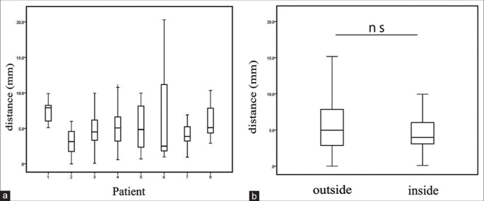
Results: Permeable films were created for a total of eight patients with intractable epilepsy. The median difference of the electrodes between the intraoperative and postoperative images was 4.6 mm (Interquartile range 2.9-7.1). The locations of electrodes implanted outside the operation field were not significantly different from those implanted inside.
Conclusion: Our new method may guide the implantation of SDEs into their planned location.
Keywords: Accuracy of the implanted electrode; Epilepsy surgery; Real-time display; Subdural electrode implantation.
Copyright: © 2024 Surgical Neurology International.
Conflict of interest statement
There are no conflicts of interest.
Figures

References
-
- Chamoun RB, Nayar VV, Yoshor D. Neuronavigation applied to epilepsy monitoring with subdural electrodes. Neurosurg Focus. 2008;25:E21. - PubMed
Grants and funding
LinkOut - more resources
Full Text Sources