. 2024 May 15;4(5):100334.
doi: 10.1016/j.bpsgos.2024.100334. eCollection 2024 Sep.

Multiple Stressors Induce Amygdalohippocampal Volume Reduction in Adult Male Rats as Detected by Longitudinal Structural Magnetic Resonance Imaging

Affiliations

Multiple Stressors Induce Amygdalohippocampal Volume Reduction in Adult Male Rats as Detected by Longitudinal Structural Magnetic Resonance Imaging

Rie Ryoke et al. Biol Psychiatry Glob Open Sci. .

Abstract

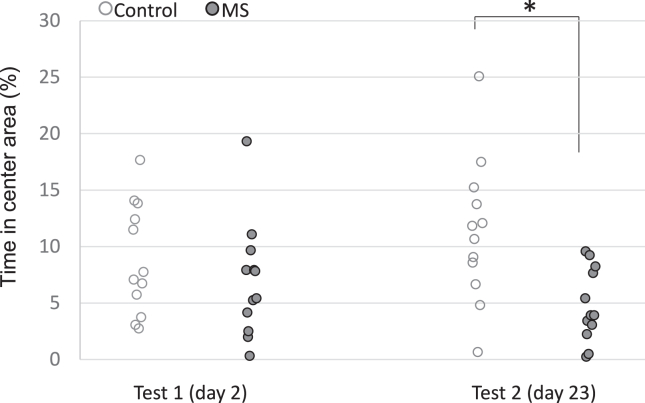
Background: Traumatic events can cause long-lasting and uncontrollable fear and anxiety. Posttraumatic stress disorder is an intractable mental disorder, and neurobiological mechanisms using animal models are expected to help development of posttraumatic stress disorder treatment. In this study, we combined multiple stress (MS) and longitudinal in vivo magnetic resonance imaging to reveal the effects of long-lasting anxiety-like behaviors on adult male rat brains.

Methods: Twelve male Wistar rats (8 weeks old) were exposed to the MS of 1-mA footshocks and forced swimming, while 12 control rats were placed in a plastic cage. Contextual fear conditioning with 0.1-mA footshocks in a context different from the MS was conducted 15 days after the MS for both groups. Three retention tests were administered after 24 hours and 9 and 16 days. Two magnetic resonance imaging scans were conducted, one on the day before MS induction and one the day after the third retention test, with a 32-day interval.

Results: The MS group showed greater freezing responses than the control group in all retention tests. Whole-brain voxel-based morphometry analyses revealed reduced gray matter volume in the anterior amygdalohippocampal area in MS group rats compared with control rats. These volume changes were negatively associated with freezing time in the third retention test in the MS group.

Conclusions: These results suggest that individual variability in the amygdalohippocampal area may be related to long-lasting fear responses after severe stress.

Keywords: Amygdala; Hippocampus; Individual variability; Magnetic resonance imaging; Posttraumatic stress disorder.

Plain language summary

Traumatic events can cause long-lasting and uncontrollable fear and anxiety. In this study, we combined multiple stress (MS) and longitudinal in vivo magnetic resonance imaging to reveal the effects of long-lasting anxiety-like behaviors on adult male rat brains. The MS group showed greater freezing responses than the control group in all retention tests. Brain morphometry analyses revealed reduced gray matter volume in the anterior amygdalohippocampal area in MS group rats compared with control rats. These results suggest that individual variability in the amygdalohippocampal area may be related to long-lasting fear responses after severe stress.


Figures

Figure 1
Figure 1
Experimental protocol. Structural MRI was performed before and after a series of behavioral tests. On day 1, the multiple stress group was exposed to multiple stressors. Both the multiple stress group and the control group engaged in the open field test on days 2 and 23 to measure anxiety-like behaviors. Both groups engaged in fear conditioning on day 15, and retention tests were conducted on days 16 (24-hour delay), 24 (9-day delay), and 31 (16-day delay). MRI, magnetic resonance imaging.
Figure 2
Figure 2
Anxiety-related reduced locomotion. Animals in the MS group showed less distance traveled than those in the control group in the open field test. While no significant group difference was observed on day 2, the MS group showed less distance traveled than the control group on day 23. ∗Significant group differences, p < .05. MS, multiple stress.
Figure 3
Figure 3
Anxiety-related exploratory behavior. Rats in the control group spent more time in the central area, showing anxiolytic and less exploratory behaviors than those in the MS group on test 2 (day 23). In contrast, the MS group showed anxiety-related exploratory behavior on test 2. ∗Significant group differences, p < .05. MS, multiple stress.
Figure 4
Figure 4
Conditioned fear responses after delays. The MS group showed exaggerated freezing compared with the control group, even after 16 days of fear conditioning. ∗Significant group difference, p < .05. MS, multiple stress.
Figure 5
Figure 5
Total volumes of brain tissues before and after treatments. There were no group differences in total gray matter, white matter, or CSF volumes on MRI 1 or 2. Brain volume in both groups were increased at MRI 2 relative to MRI 1. Error bars show standard deviations. ∗Significant group difference, p < .001. a.u., arbitrary unit; CSF, cerebrospinal fluid; MRI, magnetic resonance imaging; MS, multiple stress.
Figure 6
Figure 6
Reduced rGMV with MS associated with freezing behavior. Lower amygdalohippocampal volume in the MS group than in the control group was detected in longitudinal analyses. Familywise error–corrected p < .05 at the cluster level; uncorrected p < .001 at the voxel level. Percentage freezing time in retention test 3 of fear conditioning was negatively associated with rGMV changes in the left amygdalohippocampal area in the MS group. AP, anterior-posterior; MRI, magnetic resonance imaging; MS, multiple stress; rGMV, regional gray matter volume.

References

    1. American Psychiatric Association . Diagnostic and Statistical Manual of Mental Disorders: DSM-5TM. American Psychiatric Publishing, Inc.; Washington, DC: 2013. Posttraumatic Stress Disorder of Chapter 10. Trauma- and Stressor-Related Disorders Chapter in Section II; pp. 271–272.
    1. Charlson F., van Ommeren M., Flaxman A., Cornett J., Whiteford H., Saxena S. New WHO prevalence estimates of mental disorders in conflict settings: A systematic review and meta-analysis. Lancet. 2019;394:240–248. - PMC - PubMed
    1. Deslauriers J., Toth M., Der-Avakian A., Risbrough V.B. Current status of animal models of posttraumatic stress disorder: Behavioral and biological phenotypes, and future challenges in improving translation. Biol Psychiatry. 2018;83:895–907. - PMC - PubMed
    1. Verbitsky A., Dopfel D., Zhang N. Rodent models of post-traumatic stress disorder: Behavioral assessment. Transl Psychiatry. 2020;10:132. - PMC - PubMed
    1. Nishimura K.J., Poulos A.M., Drew M.R., Rajbhandari A.K. Know thy SEFL: Fear sensitization and its relevance to stressor-related disorders. Neurosci Biobehav Rev. 2022;142 - PMC - PubMed

LinkOut - more resources