. 2024 Jun 3;27(7):110163.
doi: 10.1016/j.isci.2024.110163. eCollection 2024 Jul 19.

MBNL2 promotes aging-related cardiac fibrosis via inhibited SUMOylation of Krüppel-like factor4

Affiliations

MBNL2 promotes aging-related cardiac fibrosis via inhibited SUMOylation of Krüppel-like factor4

Jing Lu et al. iScience. .

Abstract

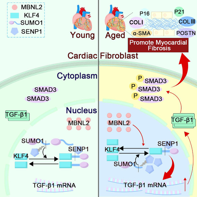
Aging-related cardiac fibrosis represents the principal pathological progression in cardiovascular aging. The Muscleblind-like splicing regulator 2 (MBNL2) has been unequivocally established as being associated with cardiovascular diseases. Nevertheless, its role in aging-related cardiac fibrosis remains unexplored. This investigation revealed an elevation of MBNL2 levels in the aged heart and senescent cardiac fibroblasts. Notably, the inhibition of MBNL2 demonstrated a capacity to mitigate H2O2-induced myofibroblast transformation and aging-related cardiac fibrosis. Further mechanistic exploration unveiled that aging heightened the expression of SENP1 and impeded the SUMO1 binding with KLF4, and SUMOylation of KLF4 effectively increased by the inhibition of MBNL2. Additionally, the inhibition of TGF-β1/SMAD3 signaling attenuated the impact of over-expression of MBNL2 in inducing senescence and cardiac fibrosis. MBNL2, by orchestrating SUMOylation of KLF4, upregulating the TGF-β1/SMAD3 signaling pathway, emerges as a significant promoter of aging-related cardiac fibrosis. This discovery identifies a novel regulatory target for managing aging-related cardiac fibrosis.

Keywords: Transcriptomics; molecular biology; omics.


Conflict of interest statement

The authors declare no competing interests.

Figures

None
Graphical abstract
Figure 1
Figure 1
RNA/ScRNA-seq database and RNA/ScRNA-seq analysis about MBNL2 (A) UMAP plots of heart disease profiles of all cells colored by cell types. (B) Bubble heatmap showing expression levels of chosen signature genes. Dot size implies a fraction of expressing cells, colored based on normalized expression levels. (C) Horizontal bar chart picturing the relative abundance of various cell types in each type sample. (D) Scatterplot of all cells in HF and N group. (E) Scatterplot of fibroblasts about HF samples in <50 and ≥50 group. (F) Correlation between MBNL2 and age from normal samples in GEO: GSE36961. (G) Volcano plot of differentially expressed gene. (H) GO enrichment analysis of DEGs.
Figure 2
Figure 2
Identification and functional verification of MBNL2 in vitro (A) Representative figure of the SA-β-gal staining. Magnification: 200×; scale bar: 50 μm. (B) Representative the immunofluorescence of α-SMA. Magnification: 200×; scale bar: 20 μm. (C) Statistical graph of SA-β-gal staining positive area. (D) Statistical graph of the immunofluorescence of α-SMA. (E and F) Representative and statistical graphs of P21, P16, COLⅠ, COLⅢ, and MBNL2 protein expression. (G) Relative mRNA level of P21, P16, COLⅠ, COLⅢ and MBNL2. (H and I) Representative and statistical graphs of the immunofluorescence of α-SMA. Magnification: 200×; scale bar: 20 μm. (J and K) Representative and statistical graphs of P21, P16, COLⅠ, COLⅢ, and MBNL2 protein. (n = 3–6, data are expressed as mean ± SEM, ∗p < 0.05, ∗∗p < 0.01 vs. the control or oe-NC group).
Figure 3
Figure 3
Functional verification of MBNL2 in vivo (A) Schematic of the experimental design. (B) Representative echocardiographic graphs. (C–J) Echocardiographic measurements of LVEF, LVFS, LVIDs, LVIDd, LVPWs, LVPWd, IVSs and IVSd. (K) Gross appearance of the whole heart. (L) H&E staining of left ventricles. Magnification: 200×; scale bar: 50 μm. (M) Masson’s staining of left ventricles. Magnification: 100×; scale bar: 100 μm. (N) HW/BW ratio of Young vs. Aged. (O) Statistical graphs of Masson staining. (P) Representative graphs of western blot of P21, P16, COLⅠ, and COLⅢ. (Q) Quantification of P21, P16, COLⅠ, and COLⅢ protein levels. (R) Relative mRNA level of P21, P16, COLⅠ and COLⅢ. (S) Representative figure of the immunofluorescence of MBNL2, Vemintin, and DAPI. Magnification: 200×; scale bar: 20 μm (T and U) Representative images and statistical analysis of IHC staining with MBNL2 antibody. Magnification: 200×; scale bar: 50 μm. (n = 3–6, data are expressed as mean ± SEM, ∗p < 0.05, ∗∗p < 0.01 vs. the young group).
Figure 4
Figure 4
Inhibition of MBNL2 mitigates aging-related cardiac fibrosis (A) Schematic of the experimental design. (B) Representative echocardiographic graphs. (C–J) Echocardiographic measurements of LVEF, LVFS, LVIDs, LVIDd, LVPWs, LVPWd, IVSs and IVSd. (K and L) H&E (Magnification: 200×; scale bar: 50 μm) and Masson’s (Magnification: 100×; scale bar: 100 μm) staining of left ventricles. (M and N) Representative images of IHC staining with α-SMA (Magnification: 100×; scale bar: 100 μm) and MBNL2 (Magnification: 200×; scale bar: 50 μm) antibody. (O) HW/BW ratio. (P) Percentage of fibrosis area. (Q and R) Statistical analysis of IHC staining with α-SMA and MBNL2 antibody. (S). Statistical analysis of MBNL2 mRNA level. (T and U) Relative western blot representative graphs and quantification of P21, P16, COLⅠ and COLⅢ protein levels. (V) Relative mRNA level of P21, P16, COLⅠ and COLⅢ. (n = 3–7, data are expressed as mean ± SEM, ∗p < 0.05, ∗∗p < 0.01 vs. the young group,#p < 0.05, ##p < 0.01 vs. the aged group, &p < 0.05, &&p < 0.01 vs. the aged+shMBNL2 group).
Figure 5
Figure 5
Effects of silencing MBNL2 on cardiac fibroblasts (A and B) Representative images and statistical graph of collagen gel contraction assay. (C–F) Representative and statistical graphs of the immunofluorescence of α-SMA and COLⅠ. Magnification: 200×; scale bar: 20 μm. (G–I) Relative mRNA levels and representative and statistical graphs of P21, P16, POSTN, COLⅠ, COLⅢ and MBNL2 protein. (n = 3–6, data are expressed as mean ± SEM, ∗p < 0.05, ∗∗p < 0.01 vs. the control group, #p < 0.05, ##p < 0.01 vs. the H2O2 group, &p < 0.05, &&p < 0.01 vs. the H2O2+sh-MBNL2 group).
Figure 6
Figure 6
Inhibition of MBNL2 activated SUMOylation of KLF4 (A–D) Representative graphs of SUMO1/2/3 protein in Young vs. Aged, Control vs. H2O2, oe-NC vs. oe-MBNL2, sh-NC vs. sh-MBNL2. (E–G) Representative and statistical graphs of SENP1 and SUMO1 protein. (H–J) Representative and statistical graphs of KLF4 and SENP1 protein. (K) Representative immunoblots of KLF4-SUMOylation after incubation with sh-MBNL2 or/and H2O2 in CFs (n = 3). (L–O) Western blot analysis and quantification of TGF-β1, T-SMAD3 and P-SMAD3 protein levels (n = 3–6, data are expressed as mean ± SEM, ∗p < 0.05, ∗∗p < 0.01 vs. the control group, #p < 0.05, ##p < 0.01 vs. the H2O2 group, &p < 0.05, &&p < 0.01 vs. the H2O2+sh-MBNL2 group).
Figure 7
Figure 7
MBNL2 inhibits fibroblast-to-myofibroblast transition through the TGF-β1/SMAD3 pathway in vitro (A and B) Representative and statistical graphs of the immunofluorescence of α-SMA. Magnification: 200×; scale bar: 20 μm. (C–F) Western blot analysis and quantification of P21, P16, POSTN, COLⅠ, COLⅢ, MBNL2, TGF-β1, T-SMAD3 and P-SMAD3 protein levels. (G) Relative mRNA level of P21, P16, POSTN, COLⅠ, COLⅢ, MBNL2 and TGF-β1 (n = 3–6, data are expressed as mean ± SEM, ∗p < 0.05, ∗∗p < 0.01 vs. the control group, #p < 0.05, ##p < 0.01 vs. the control group, &p < 0.05, &&p < 0.01 vs. the oe-MBNL2 group).

Similar articles

Cited by

References

    1. Evangelou K., Vasileiou P.V.S., Papaspyropoulos A., Hazapis O., Petty R., Demaria M., Gorgoulis V.G. Cellular senescence and cardiovascular diseases: moving to the “heart” of the problem. Physiol. Rev. 2023;103:609–647. doi: 10.1152/physrev.00007.2022. - DOI - PubMed
    1. Triposkiadis F., Xanthopoulos A., Butler J. Cardiovascular Aging and Heart Failure: JACC Review Topic of the Week. J. Am. Coll. Cardiol. 2019;74:804–813. doi: 10.1016/j.jacc.2019.06.053. - DOI - PubMed
    1. Loffredo F.S., Nikolova A.P., Pancoast J.R., Lee R.T. Heart failure with preserved ejection fraction: molecular pathways of the aging myocardium. Circ. Res. 2014;115:97–107. doi: 10.1161/CIRCRESAHA.115.302929. - DOI - PMC - PubMed
    1. Konieczny P., Stepniak-Konieczna E., Sobczak K. MBNL proteins and their target RNAs, interaction and splicing regulation. Nucleic Acids Res. 2014;42:10873–10887. doi: 10.1093/nar/gku767. - DOI - PMC - PubMed
    1. Misra C., Lin F., Kalsotra A. Deregulation of RNA Metabolism in Microsatellite Expansion Diseases. Adv. Neurobiol. 2018;20:213–238. doi: 10.1007/978-3-319-89689-2_8. - DOI - PMC - PubMed

LinkOut - more resources