

Expansion of metabolically labelled endocytic organelles and cytoskeletal cell structures in Giardia lamblia using optimised U-ExM protocols
- PMID: 38975021
- PMCID: PMC11224680
- DOI: 10.15698/mic2024.06.825
Expansion of metabolically labelled endocytic organelles and cytoskeletal cell structures in Giardia lamblia using optimised U-ExM protocols
Abstract
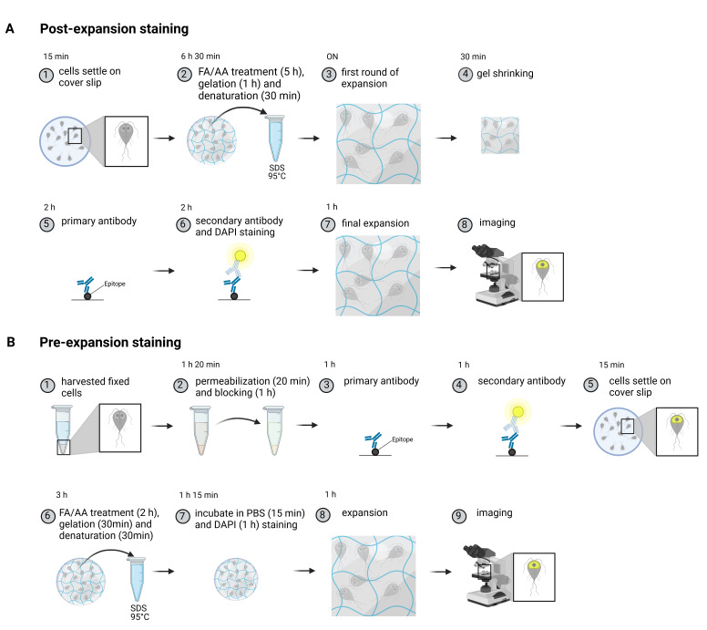
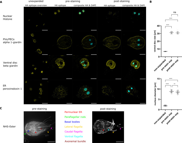
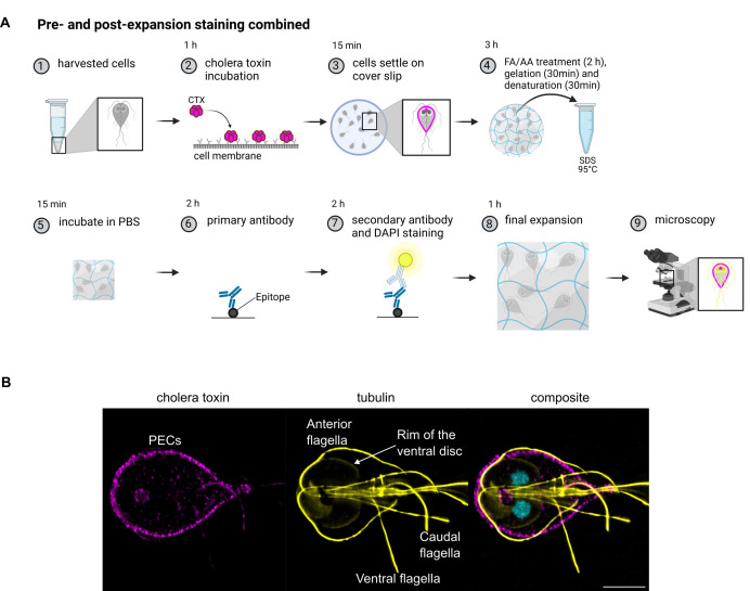
Understanding cellular ultrastructure is tightly bound to microscopic resolution and the ability to identify individual components at that resolution. Expansion microscopy has revolutionised this topic. Here we present and compare two protocols of ultrastructure expansion microscopy that allow for 4.5-fold mostly isotropic expansion and the use of antibodies, metabolic labelling, and DNA stains to demarcate individual regions such as the endoplasmic reticulum, the nuclei, the peripheral endocytic compartments as well as the ventral disc and the cytoskeleton in Giardia lamblia. We present an optimised, shortened, and modular protocol that can be swiftly adjusted to the investigators needs in this important protozoan model organism.
Keywords: Giardia lamblia; cytoskeleton; endocytosis; endoplasmic reticulum; expansion microscopy; metabolic labelling; subcellular compartment.
Conflict of interest statement
All authors declare that they have no conflicts of interest.
Figures
Similar articles
-
Ultrastructure expansion microscopy in Trypanosoma brucei.Open Biol. 2021 Oct;11(10):210132. doi: 10.1098/rsob.210132. Epub 2021 Oct 13. Open Biol. 2021. PMID: 34637654 Free PMC article.
-
Combined nanometric and phylogenetic analysis of unique endocytic compartments in Giardia lamblia sheds light on the evolution of endocytosis in Metamonada.BMC Biol. 2022 Sep 21;20(1):206. doi: 10.1186/s12915-022-01402-3. BMC Biol. 2022. PMID: 36127707 Free PMC article.
-
Clathrin-dependent pathways and the cytoskeleton network are involved in ceramide endocytosis by a parasitic protozoan, Giardia lamblia.Int J Parasitol. 2007 Jan;37(1):21-32. doi: 10.1016/j.ijpara.2006.09.008. Epub 2006 Oct 12. Int J Parasitol. 2007. PMID: 17087963 Free PMC article.
-
The structural organization of Giardia intestinalis cytoskeleton.Adv Parasitol. 2020;107:1-23. doi: 10.1016/bs.apar.2019.08.003. Epub 2019 Oct 18. Adv Parasitol. 2020. PMID: 32122527 Review.
-
Biology of Giardia lamblia.Clin Microbiol Rev. 2001 Jul;14(3):447-75. doi: 10.1128/CMR.14.3.447-475.2001. Clin Microbiol Rev. 2001. PMID: 11432808 Free PMC article. Review.
References
-
- Tillberg P W, Chen F, Piatkevich K D, Zhao Y, Yu C C, English B P, Gao L, Martorell A, Suk H J, Yoshida F, Degennaro E M, Roossien D H, Gong G, Seneviratne U, Tannenbaum S R, Desimone R, Cai D, Boyden E S. Protein-retention expansion microscopy of cells and tissues labeled using standard fluorescent proteins and antibodies. Nat Biotechnol. 2016;34(9):987–992. doi: 10.1038/nbt.3625. - DOI - PMC - PubMed
-
- Gambarotto D, Zwettler F U, Guennec M Le, Schmidt-Cernohorska M, Fortun D, Borgers S, Heine J, Schloetel JG, Reuss M, Unser M, Boyden ES, Sauer M, Hamel V, Guichard P. Imaging cellular ultrastructures using expansion microscopy (U-ExM) Nat Methods. 2018;16(1):71–74. doi: 10.1038/s41592-018-0238-1. - DOI - PMC - PubMed
LinkOut - more resources
Full Text Sources