

Expansion of metabolically labelled endocytic organelles and cytoskeletal cell structures in Giardia lamblia using optimised U-ExM protocols
- PMID: 38975021
- PMCID: PMC11224680
- DOI: 10.15698/mic2024.06.825
Expansion of metabolically labelled endocytic organelles and cytoskeletal cell structures in Giardia lamblia using optimised U-ExM protocols
Abstract
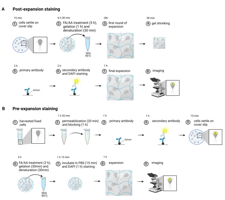
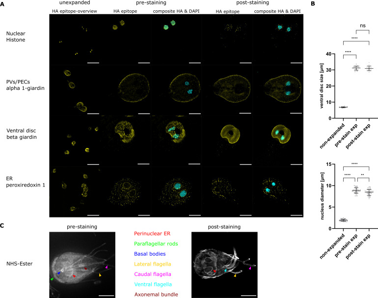
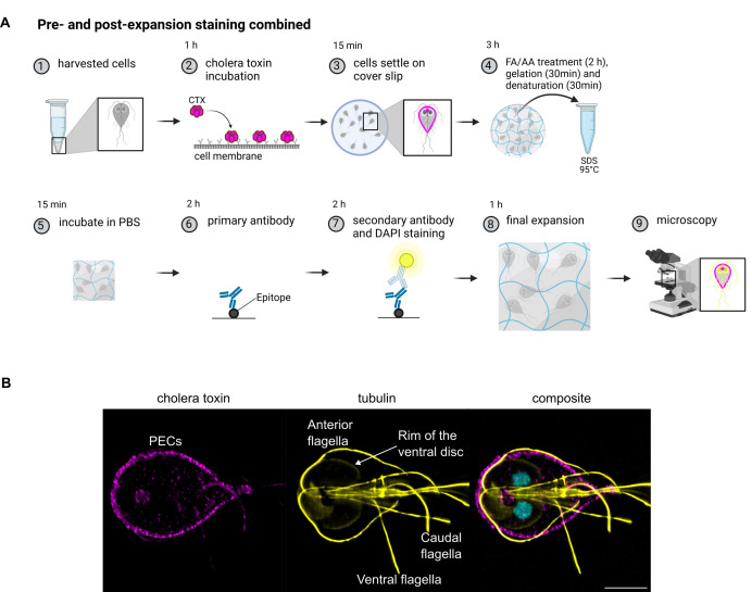
Understanding cellular ultrastructure is tightly bound to microscopic resolution and the ability to identify individual components at that resolution. Expansion microscopy has revolutionised this topic. Here we present and compare two protocols of ultrastructure expansion microscopy that allow for 4.5-fold mostly isotropic expansion and the use of antibodies, metabolic labelling, and DNA stains to demarcate individual regions such as the endoplasmic reticulum, the nuclei, the peripheral endocytic compartments as well as the ventral disc and the cytoskeleton in Giardia lamblia. We present an optimised, shortened, and modular protocol that can be swiftly adjusted to the investigators needs in this important protozoan model organism.
Keywords: Giardia lamblia; cytoskeleton; endocytosis; endoplasmic reticulum; expansion microscopy; metabolic labelling; subcellular compartment.
Conflict of interest statement
All authors declare that they have no conflicts of interest.
Figures
References
-
- Tillberg P W, Chen F, Piatkevich K D, Zhao Y, Yu C C, English B P, Gao L, Martorell A, Suk H J, Yoshida F, Degennaro E M, Roossien D H, Gong G, Seneviratne U, Tannenbaum S R, Desimone R, Cai D, Boyden E S. Protein-retention expansion microscopy of cells and tissues labeled using standard fluorescent proteins and antibodies. Nat Biotechnol. 2016;34(9):987–992. doi: 10.1038/nbt.3625. - DOI - PMC - PubMed
-
- Gambarotto D, Zwettler F U, Guennec M Le, Schmidt-Cernohorska M, Fortun D, Borgers S, Heine J, Schloetel JG, Reuss M, Unser M, Boyden ES, Sauer M, Hamel V, Guichard P. Imaging cellular ultrastructures using expansion microscopy (U-ExM) Nat Methods. 2018;16(1):71–74. doi: 10.1038/s41592-018-0238-1. - DOI - PMC - PubMed
LinkOut - more resources
Full Text Sources