. 2024 Jun 19:14:19.
doi: 10.25259/JCIS_16_2024. eCollection 2024.

Intracranial cerebrovascular lesions on T2-weighted magnetic resonance imaging

Affiliations

Intracranial cerebrovascular lesions on T2-weighted magnetic resonance imaging

Navpreet Kaur R Khurana et al. J Clin Imaging Sci. .

Abstract

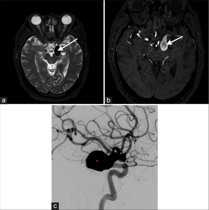
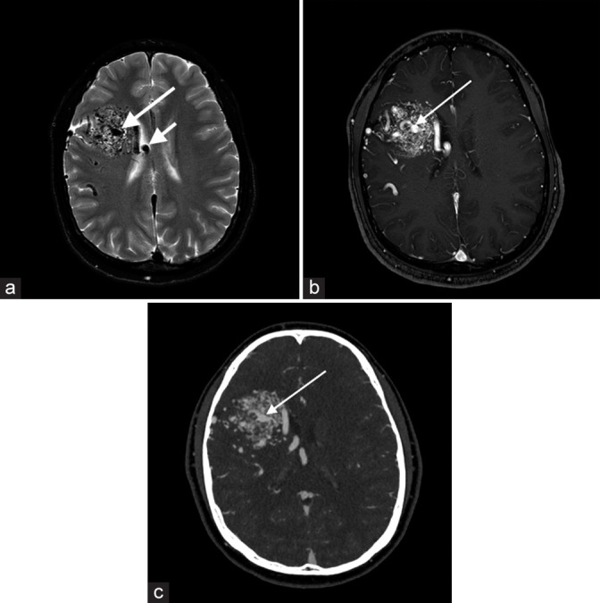
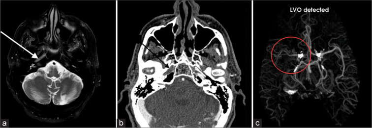
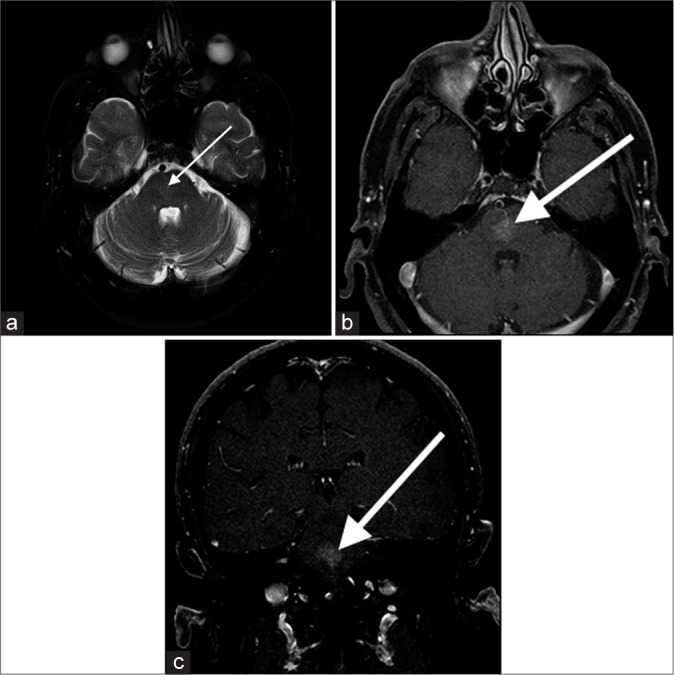
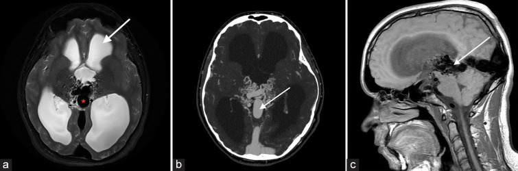
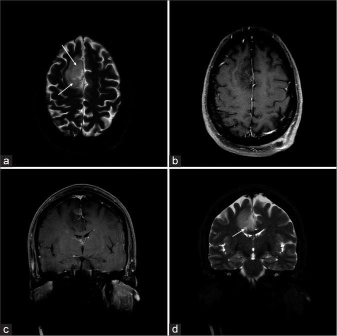
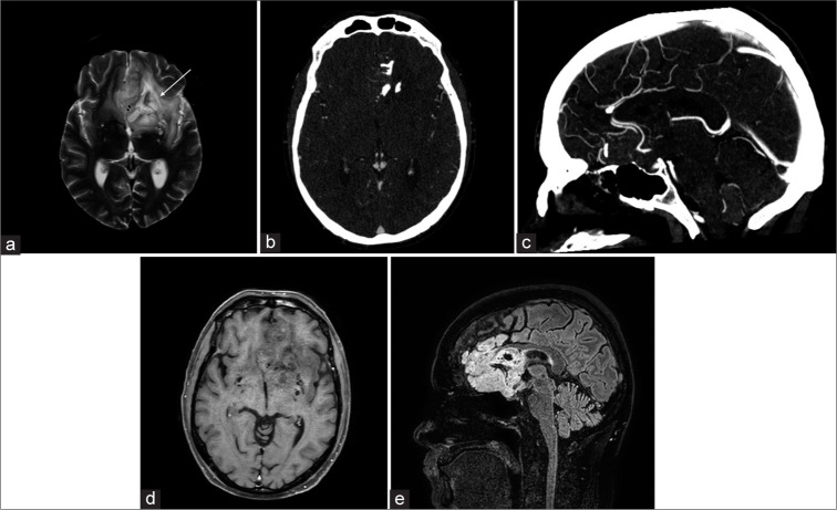
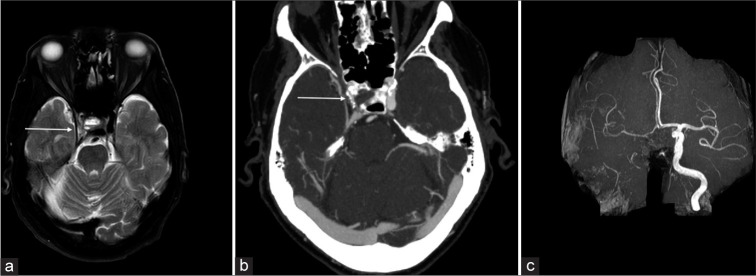
Magnetic resonance imaging (MRI) of the brain has been implemented to evaluate multiple intracranial pathologies. Non-contrast T2-weighted images are a routinely acquired sequence in almost all neuroimaging protocols. It is not uncommon to encounter various cerebrovascular lesions incidentally on brain imaging. Neuroradiologists should evaluate the routine T2-weighted images for incidental cerebrovascular lesions, irrespective of the primary indication of the study. Vascular structures typically demonstrate a low signal flow-void on the T2-weighted images. In our experience, large cerebrovascular abnormalities are easily visible to a typical neuroradiologist. In this article, we present the spectrum of the characteristic imaging appearance of various intracranial cerebrovascular lesions on routine non-contrast T2-weighted MRI. These include aneurysm, arteriovenous malformation, arterial occlusion, capillary telangiectasia, cavernous malformation, dural arteriovenous fistula, moyamoya, proliferative angiopathy, and vein of Galen malformation.

Keywords: Cerebrovascular; Incidental findings; Magnetic resonance imaging; Non-invasive imaging; Opportunistic screening.


Conflict of interest statement

There are no conflicts of interest.

Figures

Figure 1:
Figure 1:
A 68-year-old female with left carotid terminus aneurysm. (a) Axial T2-weighted image shows a lobulated T2 hypointense lesion (white arrow) projecting posteriorly from the left carotid terminus. (b) Time-of-flight magnetic resonance angiogram confirms the lesion originating from the left carotid terminus, and hence an aneurysm (white arrow). (c) Conventional catheter angiogram with the left carotid injection demonstrates opacification of the posteriorly projecting berry aneurysm (red asterisk) originating from the distal carotid artery.
Figure 2:
Figure 2:
A 20-year-old male with the right frontal arteriovenous malformation (AVM). (a) Axial T2-weighted image shows a T2 heterogeneous signal nidus (long white arrow) in the right frontal lobe with dilated adjacent T2 hypointense vessels (short white arrow). (b) Axial T1+C image demonstrates a heterogeneously enhancing lesion (long white arrow) with adjacent dilated vessels. (c) Computed tomography angiogram confirms a right frontal AVM nidus (long white arrow) with a centrally draining vein medially.
Figure 3:
Figure 3:
A 64-year-old male with the right internal carotid artery (ICA) occlusion. (a) Axial T2-weighted image shows absence of flow void (white arrow) in the petrous segment of the right ICA which is worrisome for arterial occlusion. (b) Computed tomography angiogram confirms lack of contrast related enhancement in the right petrous ICA (black arrow) consistent with occlusion. (c) Furthermore, AI detection platform detects this large vessel occlusion as well (red circle). LVO: Large vessel occlusion, AI: Artificial intelligence.
Figure 4:
Figure 4:
A 65-year-old male with pontine capillary telangiectasia. (a) Axial T2-weighted image shows a faint T2 hyperintense lesion in the pons (white arrow), and suggestive of an underlying vascular lesion. (b and c) Axial and coronal T1+C images demonstrate a faint enhancing lesion (white arrows) in the pons consistent with capillary telangiectasia.
Figure 5:
Figure 5:
A 67-year-old male with the right basal ganglia cavernous malformation. (a) Axial T2-weighted image shows a popcorn shaped lesion (white arrow) in the right posterior putamen and globus pallidum with a peripheral hypointense rim. (b) Non-contrast head computed tomography demonstrates internal calcifications (white arrow). (c) Lesion shows internal heterogeneous enhancement (white arrow) on the coronal T1+C image. Combination of these imaging findings is consistent with a cavernous malformation.
Figure 6:
Figure 6:
A 52-year-old female with the left temporal dural arteriovenous fistula (dAVF). (a) Axial T2-weighted image shows a T2 hypointense lesion (long white arrow) in the posterior left temporal lobe with adjacent dilated cortical veins (short white arrow) and surrounding edema, worrisome for an underlying vascular lesion. (b) Conventional catheter angiogram confirms Borden 3 left tentorial dAVF (red asterisk) with multiple arterial pedicles arising from the left external carotid artery (left Medial Meningeal Artery (MMA), left occipital/posterior auricular arteries). (c) Axial computed tomography angiogram image again demonstrates these findings (white arrow). MMM: Medial meningeal artery.
Figure 7:
Figure 7:
A 29-year-old female with Moyamoya. (a) Axial T2-weighted image show a cluster of vessels in the expected location of the proximal M1 segment of the left middle cerebral artery (MCA), (white arrow), and poor visualization of the carotid terminus, raising concern for underlying Moyomoya. (b) Time-of-flight magnetic resonance angiogram (MRA) confirms multiple small collateral vessels in this region. (c) 3D-rendered MRA image further confirms these findings (white arrow), with also right carotid and MCAs shown for comparison. (d) These findings are also evident on coronal maximal intensity projection computed tomography angiogram image, which also demonstrate intraventricular hemorrhage (white asterisk), which is one of the complications of Moyamoya disease. R stands for right, L stands for left.
Figure 8:
Figure 8:
A 35-year-old female with proliferative angiopathy. (a) Axial T2-weighted image shows multiple dilated T2 hypointense vessels in and along the right frontal lobe, a nidus (white arrow) and normal adjacent brain parenchyma. Findings are suggestive of a proliferative angiopathy. (b) Conventional catheter angiogram shows a larger (>3 cm) nidus (long arrow) and multiple prominent arterial feeders (short white arrow). (c) Computed tomography angiogram demonstrates abnormal vessels originating from right anterior and middle cerebral arteries (white arrow). Findings are consistent with proliferative angiopathy.
Figure 9:
Figure 9:
A 17-year-old male with vein of Galen malformation. (a) Axial T2-weighted image shows a tangled mass of abnormal connection of the arteries and the median prosencephalic vein of Markowski (white arrow). There is also associated moderate hydrocephalus (red asterisk). (b) Computed tomography angiogram demonstrates opacification of the abnormal vessels and medial prosencephalic vein (white arrow). (c) Sagittal T1-weighted image further confirms the findings (white arrow).
Figure 10:
Figure 10:
A 45-year-old male with anaplastic astrocytoma grade III, with IDH ½ mutation. (a) Axial and (b) coronal T2-weighted images show a T2 hyperintense mass centered on the left superior frontal gyrus. Internal vessels are evident as curvilinear dark flow voids (white arrows). On the (c) axial and (d) coronal T1+C images, mass is predominantly non-enhancing, with a small enhancing nodular component (asterisk). Previously seen vascular flow voids correspond to linear foci of enhancement (white arrows). IDH: Isocitrate dehydrogenase.
Figure 11:
Figure 11:
A 39-year-old male with anaplastic oligodendroglioma, with 1p/19q codeleted, IDH mutation. (a) Axial T2-weighted image shows a left frontal mass which encases the traversing bilateral anterior cerebral arteries (white arrow). (b) Axial and (c) sagittal computed tomography angiogram demonstrates characteristics internal calcifications in the mass and confirms the presence of ACAs medially. The ACA flow voids are also evident on the (d) axial T1+C and (e) sagittal FLAIR images. IDH: isocitrate dehydrogenase, ACA: Anterior cerebral artery, FLAIR: Fluid-attenuated inversion recovery.
Figure 12:
Figure 12:
A 75-year-old male with severe atherosclerotic plaque in the right internal carotid artery (ICA). (a) Axial T2-weighted image shows an asymmetrically luminal diameter of the supraclinoid segment of the right ICA (white arrow). There is also associated moderate hydrocephalus. (b) Computed tomography angiogram demonstrates severe short-segment stenosis in the cavernous and supraclinoid segments of the right ICA, with associated calcified plaque (white arrow). (c) Maximal intensity projection time-of-flight magnetic resonance angiogram image further confirms the severely diseased right ICA.

Similar articles

References

    1. Keedy A. An overview of intracranial aneurysms. Mcgill J Med. 2006;9:141–6. doi: 10.26443/mjm.v9i2.672. - DOI - PMC - PubMed
    1. Chung CY, Peterson RB, Howard BM, Zygmont ME. Imaging intracranial aneurysms in the endovascular era: Surveillance and posttreatment follow-up. Radiographics. 2022;42:789–805. doi: 10.1148/rg.210131. - DOI - PubMed
    1. Geibprasert S, Pongpech S, Jiarakongmun P, Shroff MM, Armstrong DC, Krings T. Radiologic assessment of brain arteriovenous malformations: What clinicians need to know. Radiographics. 2010;30:483–501. doi: 10.1148/rg.302095728. - DOI - PubMed
    1. Sato K, Hijikata Y, Omura N, Miki T, Kakita H, Yoshida T, et al. Usefulness of three-dimensional fast imaging employing steady-state acquisition MRI of large vessel occlusion for detecting occluded middle cerebral artery and internal carotid artery before acute mechanical thrombectomy. J Cerebrovasc Endovasc Neurosurg. 2021;23:201–9. doi: 10.7461/jcen.2021.E2020.10.007. - DOI - PMC - PubMed
    1. Lee RR, Becher MW, Benson ML, Rigamonti D. Brain capillary telangiectasia: MR imaging appearance and clinicohistopathologic findings. Radiology. 1997;205:797–805. doi: 10.1148/radiology.205.3.9393538. - DOI - PubMed

LinkOut - more resources