. 2024 Jun 11;10(12):e32730.
doi: 10.1016/j.heliyon.2024.e32730. eCollection 2024 Jun 30.

Interferon-α could induce liver steatosis to promote HBsAg loss by increasing triglyceride level

Affiliations

Interferon-α could induce liver steatosis to promote HBsAg loss by increasing triglyceride level

Lili Wu et al. Heliyon. .

Abstract

Background: The correlation between metabolic syndrome (MetS) and hepatitis B surface antigen (HBsAg) loss remains to be further elucidated, particularly in patients receiving pegylated interferon-α (PEG-IFN) treatment.

Methods: 758 patients with low HBsAg quantification who had received nucleos(t)ide analog (NUC) therapy for at least one year and subsequently switched to or add on PEG-IFN therapy over an unfixed course were enrolled. 412 patients were obtained with baseline data matched. A total of 206 patients achieved HBsAg loss (cured group) within 48 weeks. Demographic and biochemical data associated with MetS were gathered for analysis. HepG2.2.15 cell line was used in vitro experiments to validate the efficacy of interferon-α (IFN-α).

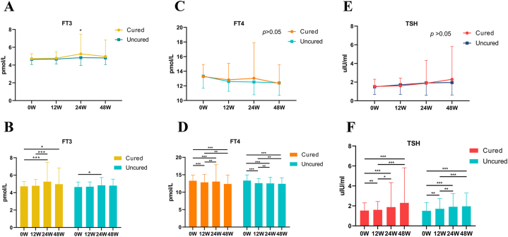
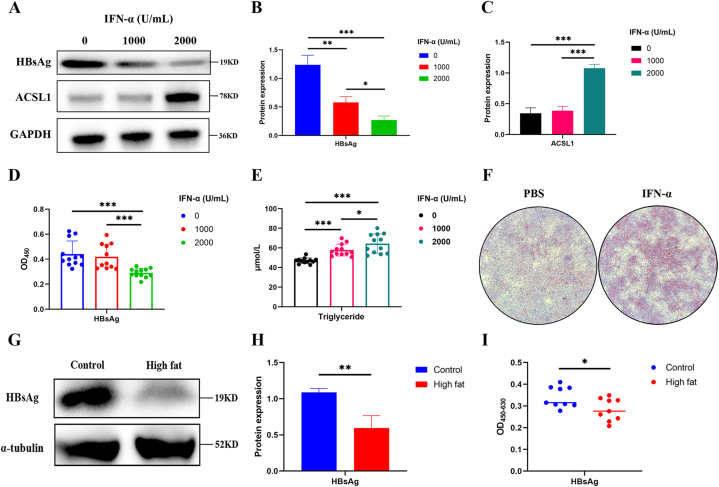
Results: The proportion of patients with diabetes or hypertension in the uncured group was significantly higher than in the cured group. The levels of fasting blood glucose (FBG) and glycated albumin remained elevated in the uncured group over the 48 weeks. In contrast, the levels of blood lipids and uric acid remained higher in the cured group within 48 weeks. Triglycerides levels and liver steatosis of all patients increased after PEG-IFN therapy. Baseline elevated uric acid levels and hepatic steatosis may be beneficial for HBsAg loss. IFN-α could induce hepatic steatosis and indirectly promote HBsAg loss by increasing triglyceride level through upregulation of acyl-CoA synthetase long-chain family member 1(ACSL1).

Conclusions: IFN-α could induce liver steatosis to promote HBsAg loss by increasing triglyceride level through upregulation of ACSL1. Comorbid diabetes may be detrimental to obtaining HBsAg loss with PEG-IFN therapy in CHB patients.

Keywords: ACSL1; Chronic hepatitis B; HBsAg loss; Interferon-alfa; Liver steatosis; Triglyceride.


Conflict of interest statement

The authors declare that they have no known competing financial interests or personal relationships that could have appeared to influence the work reported in this paper.

Figures

Fig. 1
Fig. 1
Patient enrollment.
Fig. 2
Fig. 2
Differences in medication regimens and comorbidities between the two groups of patients. Medication regimens for the two groups of patients (A–B). The proportion of patients with type 2 diabetes (C) or hypertension (D) in the two groups. The proportion of patients with a history of alcohol consumption (E) and family history (F) in the two groups. *p < 0.05, **p < 0.01, ***p < 0.001, “ns” indicates no significance. PEG-IFN, pegylated interferon-α; ETV: entecavir; TDF, tenofovir disoproxil fumarate; TAF, tenofovir alafenamide; ADV, adefovir dipivoxil; LAM, lamivudine.
Fig. 3
Fig. 3
Difference in blood glucose between the two groups. The difference and the dynamic changes in fasting blood glucose (A–B) and glycated albumin (C–D) levels between the cured and uncured patients at different time points. “ns” indicates no significance, *p < 0.05, **p < 0.01, ***p < 0.001.
Fig. 4
Fig. 4
Differences in lipids and uric acid between the two groups of patients. The difference and dynamic changes in total cholesterol (CHOL) (A–B), high-density lipoprotein (HDL) (C–D), low-density lipoprotein (LDL) (E–F), triglyceride (TRIG) (G–H) and uric acid (UA) (I–J) levels between cured and uncured patients at different time points. *p < 0.05, **p < 0.01, ***p < 0.001.
Fig. 5
Fig. 5
Differences in the level of hepatic steatosis as well as fibrosis between the two groups of patients. The difference and dynamic changes in body mass index (BMI) (A–B), controlled attenuation parameter (CAP) (C–D) and liver stiffness measurement (LSM) (E–F) levels between cured and uncured patients at different time points. “ns” indicates no significance, *p < 0.05, **p < 0.01, ***p < 0.001.
Fig. 6
Fig. 6
Increased hepatic steatosis after interferon-α therapy. Differences in liver steatosis grading before and after PEG IFN treatment in two groups of patients (A–B). The difference and dynamic changes in the proportion of liver steatosis diagnosed by ultrasound (C–D) between cured and uncured patients at different time points. The baseline uric acid (UA) level (E) and controlled attenuation parameter (CAP) value (F) were negatively correlated with HBsAg quantification.
Fig. 7
Fig. 7
Differences in thyroid function between the two groups. The difference and dynamic changes in free triiodothyronine (FT3) (A–B), free thyroxine (FT4) (C–D) and thyroid stimulating hormone (TSH) (E–F) levels between cured and uncured patients at different time points. *p < 0.05, **p < 0.01, ***p < 0.001.
Fig. 8
Fig. 8
IFN-α could induce liver steatosis to promote HBsAg loss by increasing triglyceride level through upregulation of ACSL1. IFN-α significantly down-regulated intracellular and extracellular HBsAg levels and up-regulated ACSL1 expression (A–D). IFN-α treatment upregulated intra- and extracellular triglyceride levels (E–F). The intracellular and extracellular HBsAg levels could be down-regulated by high-fat medium conditions compared to normal medium (G–I). IFN-α: interferon-alfa; ACSL1: acyl-CoA synthetase long-chain family member 1; HBsAg: hepatitis B surface antigen. “ns” indicates no significance, *p < 0.05, **p < 0.01, ***p < 0.001.

References

    1. Revill P.A., Chisari F.V., Block J.M., et al. A global scientific strategy to cure hepatitis B. Lancet Gastroenterol Hepatol. 2019;4(7):545–558. doi: 10.1016/s2468-1253(19)30119-0. - DOI - PMC - PubMed
    1. Terrault N.A., Lok A.S.F., McMahon B.J., et al. Update on prevention, diagnosis, and treatment of chronic hepatitis B: AASLD 2018 hepatitis B guidance. Hepatology. 2018;67(4):1560–1599. doi: 10.1002/hep.29800. - DOI - PMC - PubMed
    1. Ning Q., Wu D., Wang G.Q., et al. Roadmap to functional cure of chronic hepatitis B: an expert consensus. J. Viral Hepat. 2019;26(10):1146–1155. doi: 10.1111/jvh.13126. - DOI - PubMed
    1. Ning Q., Han M., Sun Y., et al. Switching from entecavir to PegIFN alfa-2a in patients with HBeAg-positive chronic hepatitis B: a randomised open-label trial (OSST trial) J. Hepatol. 2014;61(4):777–784. doi: 10.1016/j.jhep.2014.05.044. - DOI - PubMed
    1. Wu D., Wang P., Han M., et al. Sequential combination therapy with interferon, interleukin-2 and therapeutic vaccine in entecavir-suppressed chronic hepatitis B patients: the Endeavor study. Hepatol Int. 2019;13(5):573–586. doi: 10.1007/s12072-019-09956-1. - DOI - PubMed

LinkOut - more resources