. 2024 Jul 8;22(7):e3002684.
doi: 10.1371/journal.pbio.3002684. eCollection 2024 Jul.

The new science of sleep: From cells to large-scale societies

Affiliations

The new science of sleep: From cells to large-scale societies

Omer Sharon et al. PLoS Biol. .

Abstract

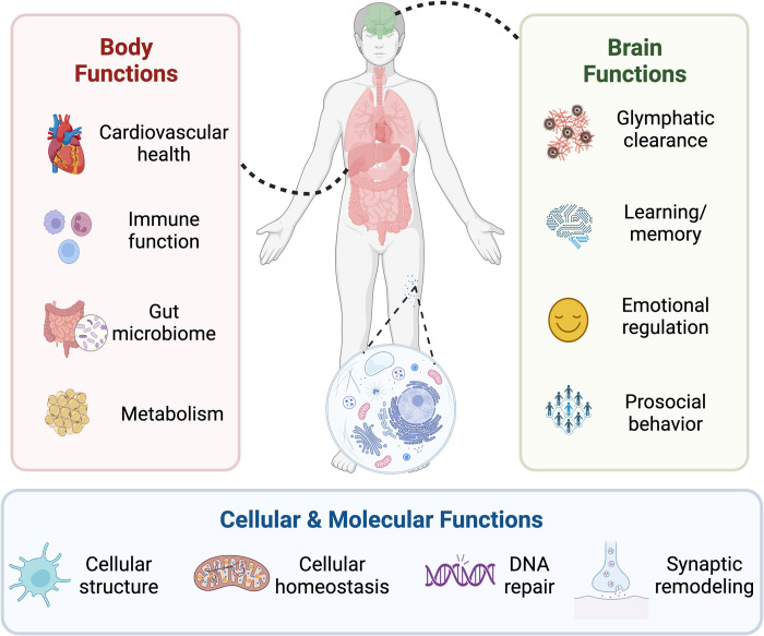
In the past 20 years, more remarkable revelations about sleep and its varied functions have arguably been made than in the previous 200. Building on this swell of recent findings, this essay provides a broad sampling of selected research highlights across genetic, molecular, cellular, and physiological systems within the body, networks within the brain, and large-scale social dynamics. Based on this raft of exciting new discoveries, we have come to realize that sleep, in this moment of its evolution, is very much polyfunctional (rather than monofunctional), yet polyfunctional for reasons we had never previously considered. Moreover, these new polyfunctional insights powerfully reaffirm sleep as a critical biological, and thus health-sustaining, requisite. Indeed, perhaps the only thing more impressive than the unanticipated nature of these newly emerging sleep functions is their striking divergence, from operations of molecular mechanisms inside cells to entire group societal dynamics.


Conflict of interest statement

The authors have declared that no competing interests exist.

Figures

Fig 1
Fig 1. The necessity of sleep at multiple levels.
Sleep serves a multitude of functions for humans. These functions exist at multiple physiological levels, from cells (bottom panel) to bodily systems (left panel), through to multiple brain functions and systems (right panel).
Fig 2
Fig 2. Artificially boosting sleep.
Several different noninvasive methods have been developed for artificially augmenting human sleep. (A) Electrical brain stimulation, including when it is time-locked to the upcoming peaks of individual deep NREM sleep brain waves, can enhance the power of those slow waves in a mechanism similar to the external assistance, or pushing, of a swing. (B) A similar outcome can also be achieved by slowly rocking a bed at frequencies close to the slowest oscillations of deep NREM sleep (purple, approximately 0.25 Hz), leading to an increase in the amount of deep sleep, and helping with a faster sleep onset, relative to a stationary bed (gray). (C) Thermal stimulation of specific regions of the body represents another method for artificially improving human sleep. Normally, the mechanism instigating human sleep (sleep onset) involves an increase in skin peripheral temperature of vascular regions such as the hands and feet (yellow dashed line). As the blood rises to the surface away from the inner body, core body temperature decreases, and the coincidence of these 2 changes provides a thermal signal triggering sleep onset (red dashed line, left-side panel). Thereafter, further decreasing core body temperature is associated with increasing amounts of deep NREM sleep. By artificially accelerating these transitions, mostly by experimentally warming the hands and feet, core body temperature decreases more rapidly, therefore reducing the time it takes those individuals to fall asleep (right-side panel), with further such thermal intervention subsequently increasing the amount of deep NREM sleep and reducing the amount of nighttime awakenings (i.e., increasing sleep stability and the consolidated nature of sleep).
Fig 3
Fig 3. Social and emotional consequences of sleep loss.
(A) Within an individual, sleep loss (pink) triggers a sharp reduction in positive mood and, to a lesser extent, an increase in negative mood (left-side panel). The emotional intensity felt by sleep-deprived individuals is also amplified by lack of sleep. However, there is a paradoxical decrease in the outward emotional expressivity triggered by sleep deprivation (right-side panel). These affective changes are further reflected in the brain. Here, sleep loss increases activity in the limbic network involved in emotional processing (red, left-side brain) yet reduces activity in the mPFC (blue, right-side brain). In addition, functional connectivity between the mPFC and amygdala is also reduced by sleep loss (dashed blue line), which is a communication pathway that normally regulates emotion. (B) Interindividual affective processes and behaviors are also altered by sleep loss. For example, sleep loss increases feelings of loneliness within the sleep-deprived individual and lowers feelings of empathy towards others (left panel). This asocial phenotype within an individual is further reflected in the reduced desire to interact with other, rested individuals. This effect is bidirectional. Rested individuals, unknowing of the sleep-deprived state of their conspecific, nevertheless show a similar reduction in the desire to interact with underslept others (right panel). (C) Across larger societal scales, insufficient sleep impairs prosocial behavior observed in large groups of individuals. For example, underslept groups express a reduced overall trend of helping behaviors and reduced motivation of typical societal civic duties, such as volunteering or voting (left panel). One underlying mechanism accounting for these collective asocial consequences is impaired activity in the social cognition brain network of underslept individuals (right panel), which is relevant as this network normally supports the ability to understand the state of others (i.e., theory of mind), and also promotes prosocial helping and cooperation. ACC, anterior cingulate cortex; mPFC, medial prefrontal cortex; TPJ, temporal parietal junction.
Fig 4
Fig 4. Dreaming and the brain.
(A) Brain activation during REM sleep—one of the principal stages associated with vivid dreaming. Relative to brain activity when an individual is either awake or in non-REM sleep, there is increased activation of visual, sensorimotor, and affective pathways during REM sleep (golden clusters). Additional regions then come online and are activated when individuals experience lucid REM sleep (red clusters; relative to nonlucid REM sleep). These include regions of the anterior prefrontal cortex involved in volitional executive decisions and actions, and the precuneus, involved in self-referential processing. (B) Incorporation of recent waking events into dreams unfolds in a 2-peak reliable pattern over time. The first temporal peak of waking incorporations occurs on the first 2 nights and then fades. However, these same prior waking experiences reemerge as a second peak 5–7 days later. This temporal pattern of waking life incorporation is known as the dream lag effect. (C) IRT is a behavioral intervention method for treating and dissipating nightmares. IRT includes the waking rehearsal of alternatives to nightmare scenarios, developed between the patient and their therapist. These more neutral or positive alternatives to the nightmare scenario are rehearsed daily by the patient for up to 2 weeks. As a result, the nightmares become significantly less distressing. A recent study added an additional methodological step. During the daytime rehearsal of the nightmare alternative, an auditory tone (here, a piano chord) was played every 10 seconds in the background. Then, as the patient slept and went into REM sleep—the stage most commonly associated with nightmares—the same piano chord was played at a level that did not wake the patient up. The goal was to reactivate the memory of the alternative scenario as the sleeping brain is processing. As a result, patients experienced an even larger decrease in the distressing nature of the nightmare, relative to standard IRT. IRT, imagery rehearsal therapy; REM, rapid eye movement.
Fig 5
Fig 5. Sleep variability across species.
Sleep duration varies markedly across the entire animal kingdom, from invertebrates to mammals. In most invertebrates and fish, sleep is defined behaviorally (e.g., for fire ants [276] and Port Jackson sharks [277]). Physiological evidence of NREM sleep can be found in a few amphibian species (e.g., the common frog [278]), as well as in reptiles [279]. In birds and mammals, evidence of sleep includes physiological recordings of both REM and NREM sleep, often measured in the lab [–282]. NREM, nonrapid eye movement; REM, rapid eye movement.

References

    1. Walker MP. Sleep essentialism. Brain. 2021;144:697–699. doi: 10.1093/brain/awab026 - DOI - PubMed
    1. Lesku JA, Schmidt MH. Energetic costs and benefits of sleep. Curr Biol. 2022;32:R656–R661. doi: 10.1016/j.cub.2022.04.004 - DOI - PubMed
    1. Czeisler CA. Duration, timing and quality of sleep are each vital for health, performance and safety. Sleep Health. 2015;1:5–8. doi: 10.1016/j.sleh.2014.12.008 - DOI - PubMed
    1. Chattu VK, Manzar MD, Kumary S, Burman D, Spence DW, Pandi-Perumal SR. The Global Problem of Insufficient Sleep and Its Serious Public Health Implications. Healthcare (Basel). 2018:7. doi: 10.3390/healthcare7010001 - DOI - PMC - PubMed
    1. He Y, Jones CR, Fujiki N, Xu Y, Guo B, Holder JL Jr, et al. The transcriptional repressor DEC2 regulates sleep length in mammals. Science. 2009;325:866–870. doi: 10.1126/science.1174443 - DOI - PMC - PubMed